Copyright
©The Author(s) 2024.
World J Clin Cases. Oct 6, 2024; 12(28): 6180-6186
Published online Oct 6, 2024. doi: 10.12998/wjcc.v12.i28.6180
Published online Oct 6, 2024. doi: 10.12998/wjcc.v12.i28.6180
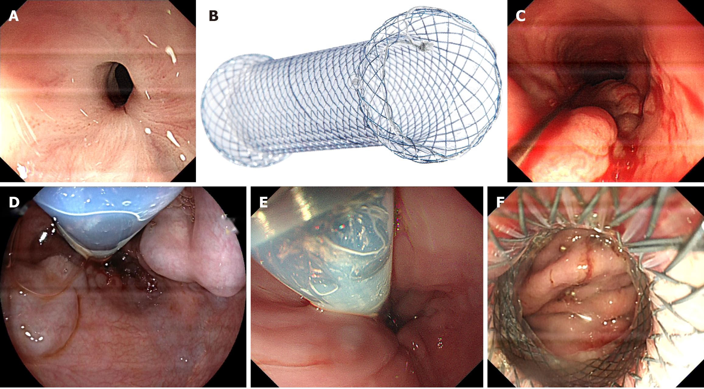
Figure 1 Observing the retention in the esophageal cavity using an endoscope, and we should be alert to residual varices.
A: The stricture was exposed under the endoscope; B: This is a semi covered self-expanding metal stent; C: The self-expanding metal stent was passed through the stricture by a hard wire guarding; D: We should be alert to the residual varices, when we were placing the self-expanding metal stent; E: The oral side of stent should be above and near to the stricture; F: The anal side of stent should be passed cardia.
Figure 2 The position of stent.
A: After expanding, the oral side of stent was well; B: After expanding, the endoscope was passed through the stent; C: After expanding, the endoscope was passed through stent into the stomach to confirm that anus of stent being well.
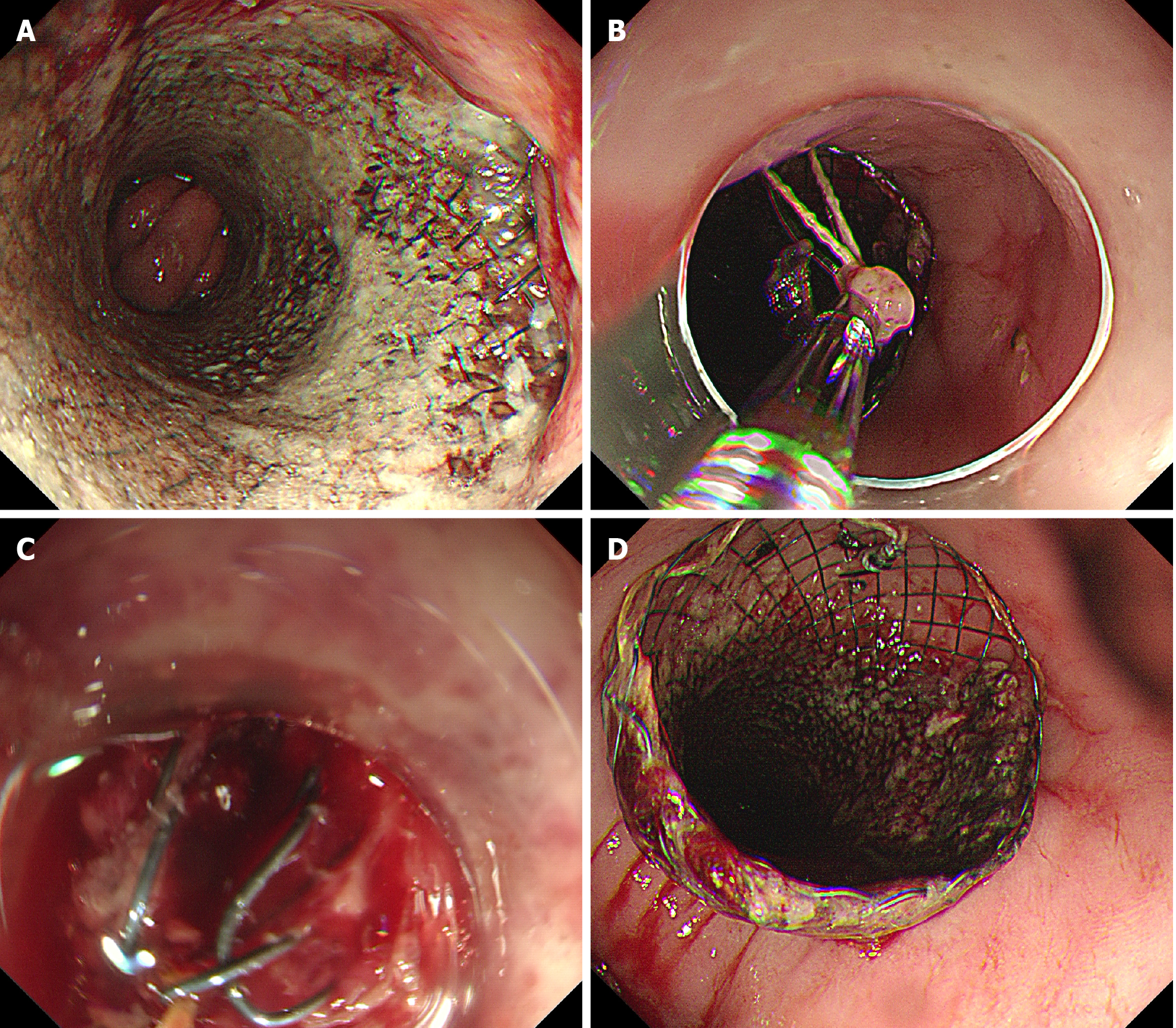
Figure 3 Clamping the knot of stent and pull with forceps to adjust the self-expanding metal stent if the stent embedd into esophageal mucosa after placing.
A: After placing for one month, the stent embedd into esophageal mucosa; B: The knot of stent was clamped and pulled with forceps to adjust the self-expanding metal stent; C: The stent was being adjusted with the forceps; D: After adjusting, the position of self-expanding metal stent was well.
- Citation: Zhang FL, Xu J, Jiang YH, Zhu YD, Shi Y, Li X, Wang H, Huang CJ, Zhou CH, Zhu Q, Chen JW. Self-expanding metal stent for relieving the stricture after endoscopic injection for esophageal varices. World J Clin Cases 2024; 12(28): 6180-6186
- URL: https://www.wjgnet.com/2307-8960/full/v12/i28/6180.htm
- DOI: https://dx.doi.org/10.12998/wjcc.v12.i28.6180


