Copyright
©The Author(s) 2024.
World J Clin Cases. Sep 26, 2024; 12(27): 6117-6123
Published online Sep 26, 2024. doi: 10.12998/wjcc.v12.i27.6117
Published online Sep 26, 2024. doi: 10.12998/wjcc.v12.i27.6117
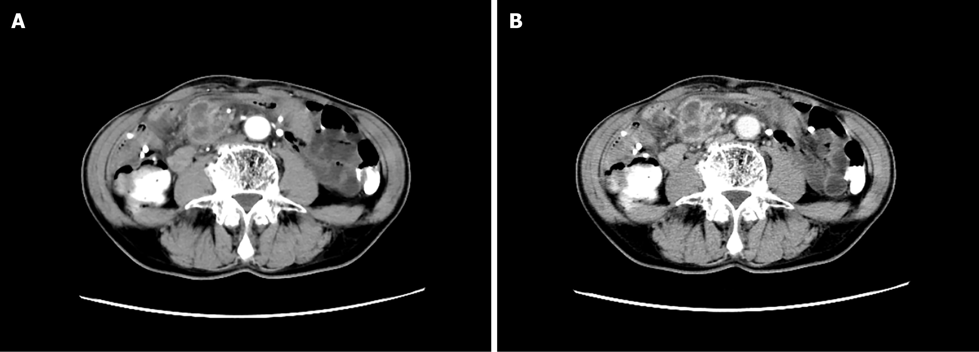
Figure 1 Enhanced computed tomography.
A: Arterial phase; B: Venous phase. They showed a right lower abdominal mass with scattered multiple nodules in the abdominal cavity.
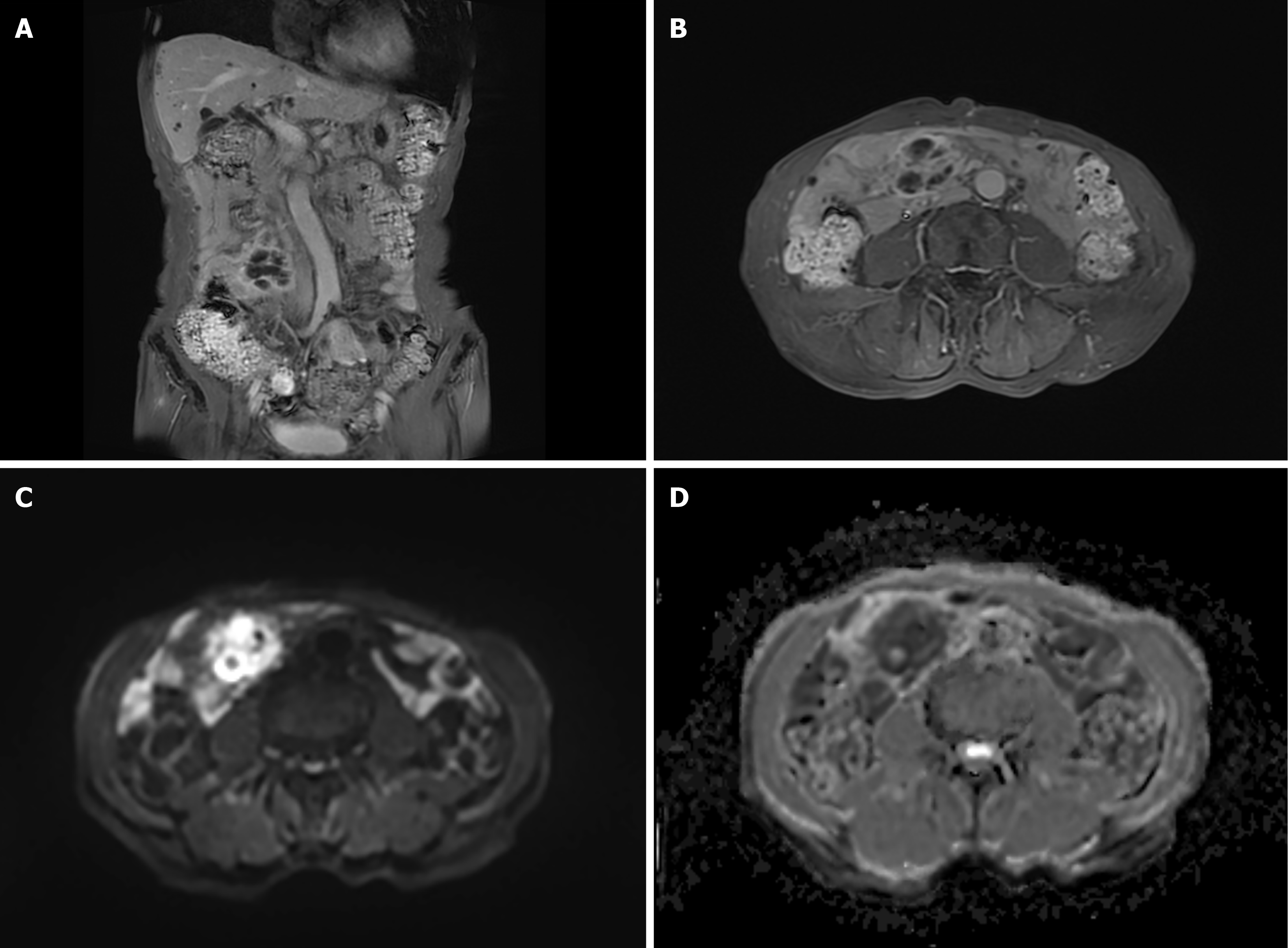
Figure 2 Different sequences of magnetic resonance dynamic enhancement + diffusion-weighted imaging of the lower abdomen.
A: T1_quick3d_cor_fs_bh; B: T1_quick3d_tra_fs_bh; C: Epi_dwi_tra_trig_b800; D: Epi_dwi_tra_trig_ADC. They showed abnormally enhanced foci of the right lower abdominal cavity of an undetermined nature.
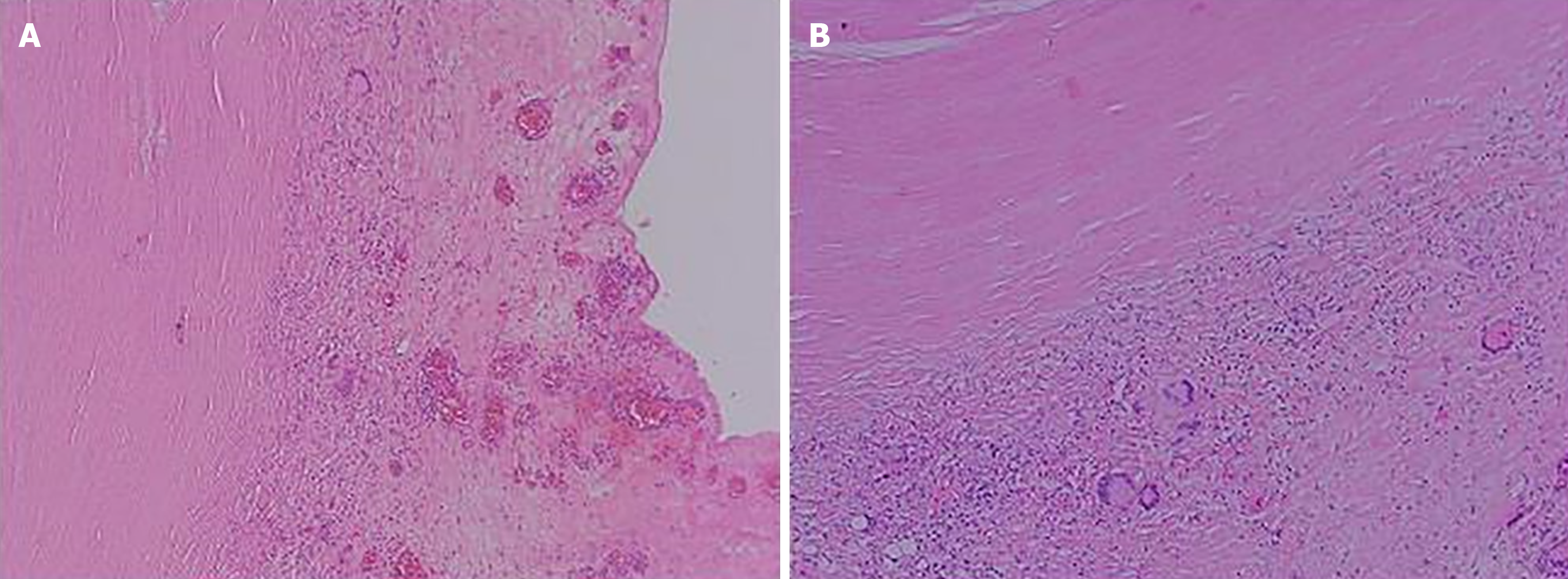
Figure 3 Postoperative pathology.
A: Abdominal mass; B: Intra-abdominal nodule. They showed peripheral fibrotic tissue proliferation with multinucleated giant cell reaction (hematoxylin and eosin, 100 ×).
- Citation: Liu WP, Ma FZ, Zhao Z, Li ZR, Hu BG, Yang T. Tuberculous peritonitis complicated by an intraperitoneal tuberculous abscess: A case report. World J Clin Cases 2024; 12(27): 6117-6123
- URL: https://www.wjgnet.com/2307-8960/full/v12/i27/6117.htm
- DOI: https://dx.doi.org/10.12998/wjcc.v12.i27.6117


