Copyright
©The Author(s) 2024.
World J Clin Cases. Sep 16, 2024; 12(26): 5983-5989
Published online Sep 16, 2024. doi: 10.12998/wjcc.v12.i26.5983
Published online Sep 16, 2024. doi: 10.12998/wjcc.v12.i26.5983
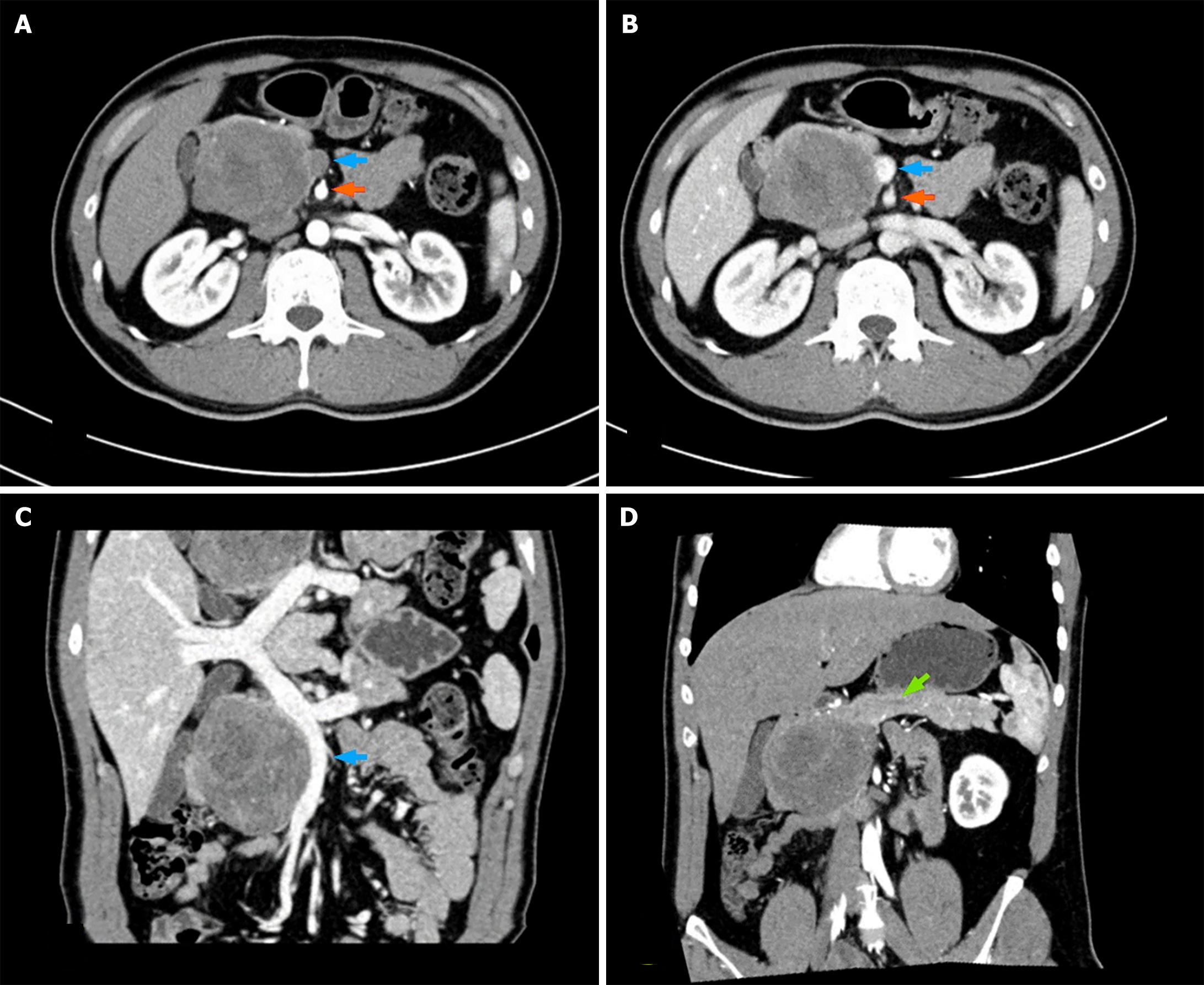
Figure 1 An enhanced computed tomography image of the abdomen revealed a mass in the head of the pancreas.
A: Arterial phase; B: Portal phase; C: Portal venous system reconstruction; D: Coronal reconstruction. Blue arrow: Superior mesenteric vein; Orange arrow: Superior mesenteric artery; Green arrow: Main pancreatic duct.
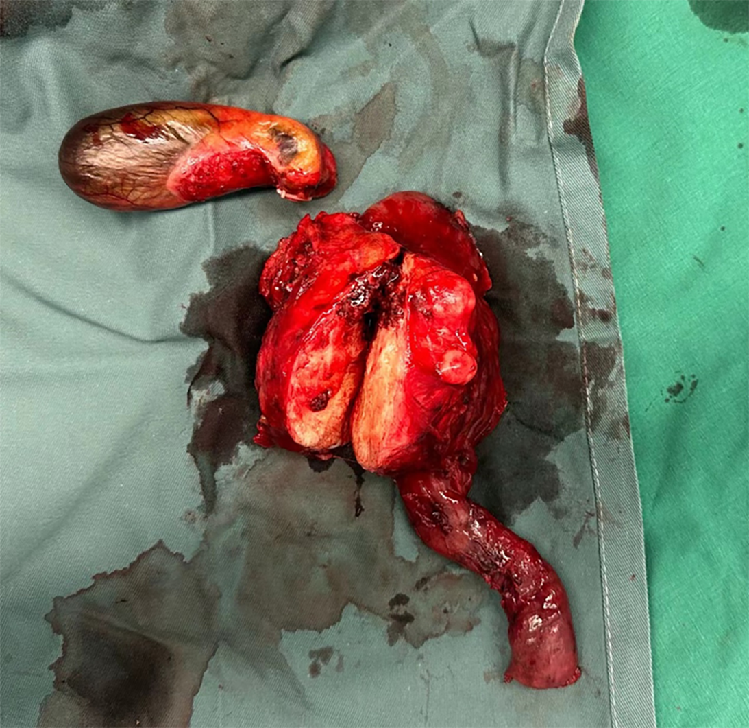
Figure 2 Macroscopic photographs of the resected specimen.
The tumor was solid, gray-white, hard in texture, and had roughly clear borders.
Figure 3 Histopathological sections of the resected specimen.
A: Low × 10 and medium × 20 (inserted) magnification H and E stains demonstrating that the tumor cells are epithelioid and that mitosis is easily observed; B: Mucin 4 stain demonstrating strong and diffuse staining of the tumor cells.
- Citation: Sun MQ, Guo LN, You Y, Qiu YY, He XD, Han XL. Sclerosing epithelioid fibrosarcoma of the pancreas: A case report. World J Clin Cases 2024; 12(26): 5983-5989
- URL: https://www.wjgnet.com/2307-8960/full/v12/i26/5983.htm
- DOI: https://dx.doi.org/10.12998/wjcc.v12.i26.5983


