Copyright
©The Author(s) 2024.
World J Clin Cases. Sep 16, 2024; 12(26): 5952-5959
Published online Sep 16, 2024. doi: 10.12998/wjcc.v12.i26.5952
Published online Sep 16, 2024. doi: 10.12998/wjcc.v12.i26.5952
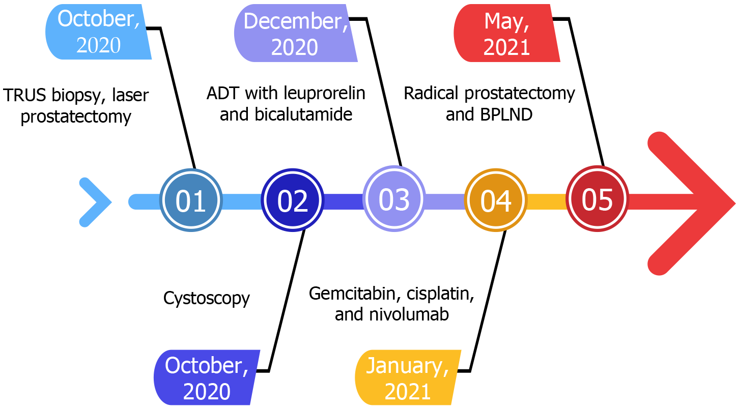
Figure 1 Fishbone diagram of treatment.
ADT: Androgen deprivation therapy; BPLND: Bilateral pelvic lymph node dissection; TRUS: Transrectal ultrasound.
Figure 2 Image of the pelvis obtained during follow-up with magnetic resonance imaging.
A: A 2.4-cm lesion with low T2 signal is observed in the posterolateral portion of the left transitional zone; B: Apparent diffusion coefficient of the lesion with low signal; C: Increased diffusion tensor imaging signals are observed.
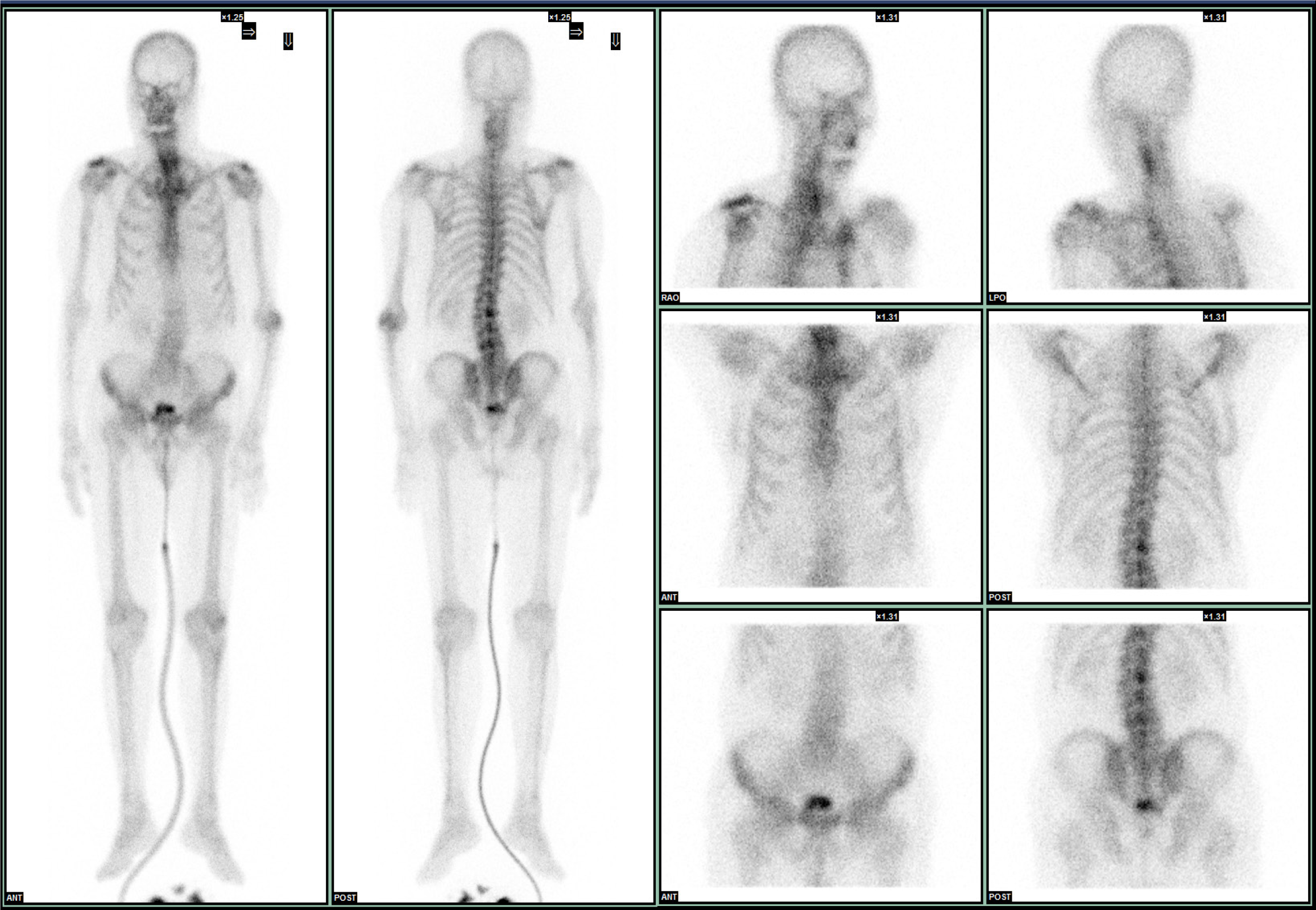
Figure 3 Technetium-99m methylene diphosphonate whole-body bone scan.
Focal areas of increased radioactivity uptake are noted in the lumbar spine at levels L1/2, L3/4, and L4/5, as well as in the left elbow and bilateral acromioclavicular joints. These findings suggest a benign etiology.
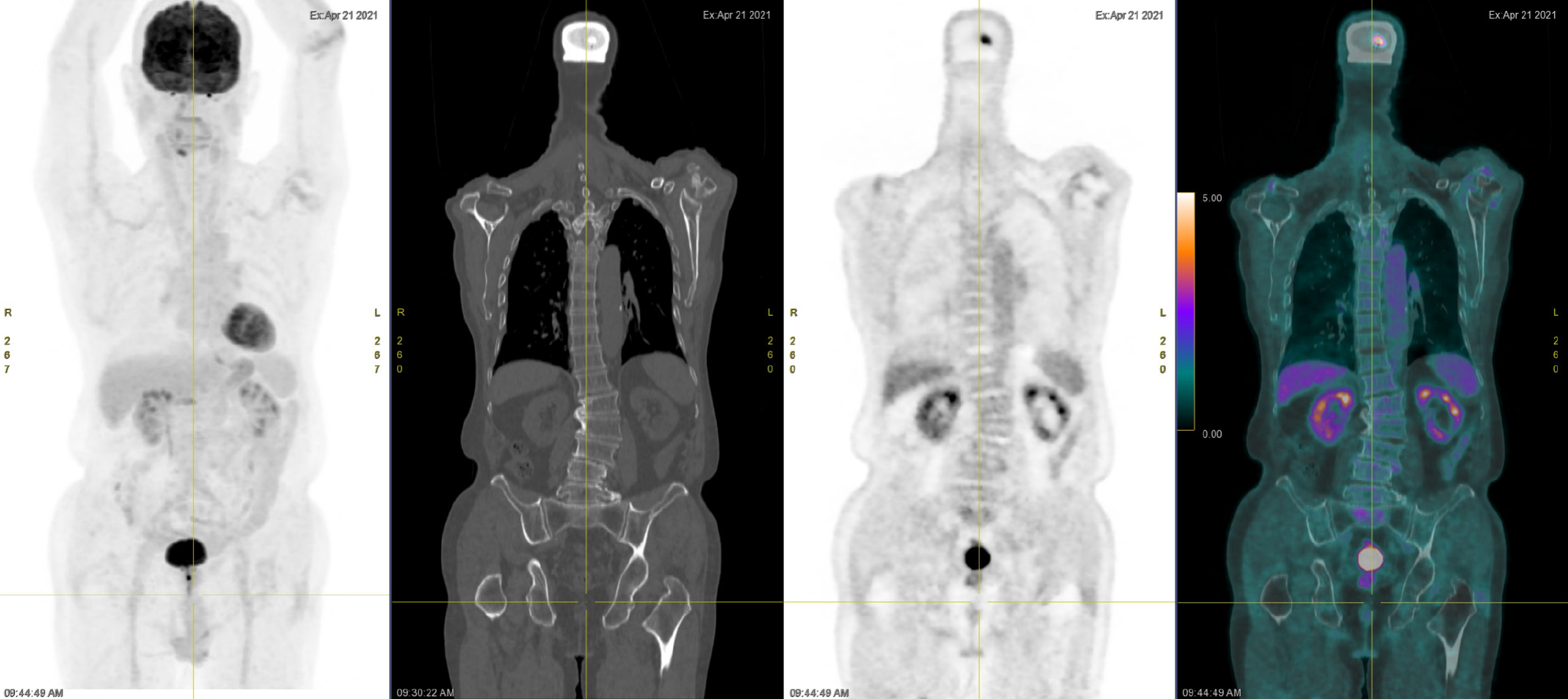
Figure 4 Images obtained during an 18F-fluorodeoxyglucose positron emission tomography evaluation of the region comprising the head to the upper thigh.
From left to right: Ill-defined fluorodeoxyglucose (FDG) avidity is observed in the prostate; No FDG-avid regional or distant lymph nodes are apparent; Ill-defined FDG avidity is noted in the right buccal region after the dental prosthesis; and Ill-defined FDG avidity in the right T1, T4, T8, T12, and left upper ilia with sclerotic changes, suggesting a benign etiology.
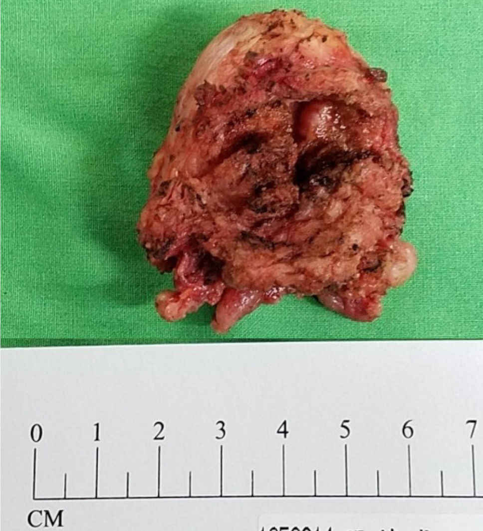
Figure 5 Gross examination of the prostate tumor after robotic-assisted radical prostatectomy.
4 cm × 3 cm × 4 cm prostate gland with a volume of 24.96 mL and weight of 29 g; The 19 cm × 1.3 cm × 0.7 cm adenocarcinoma is located in the left anterior lateral lobe and, specifically, in the middle area transitioning to the peripheral zone. The 1 cm × 0.6 cm × 0.6 cm carcinoma of the urethra is observed in the right posterior lobe, middle area, and near the periurethra.
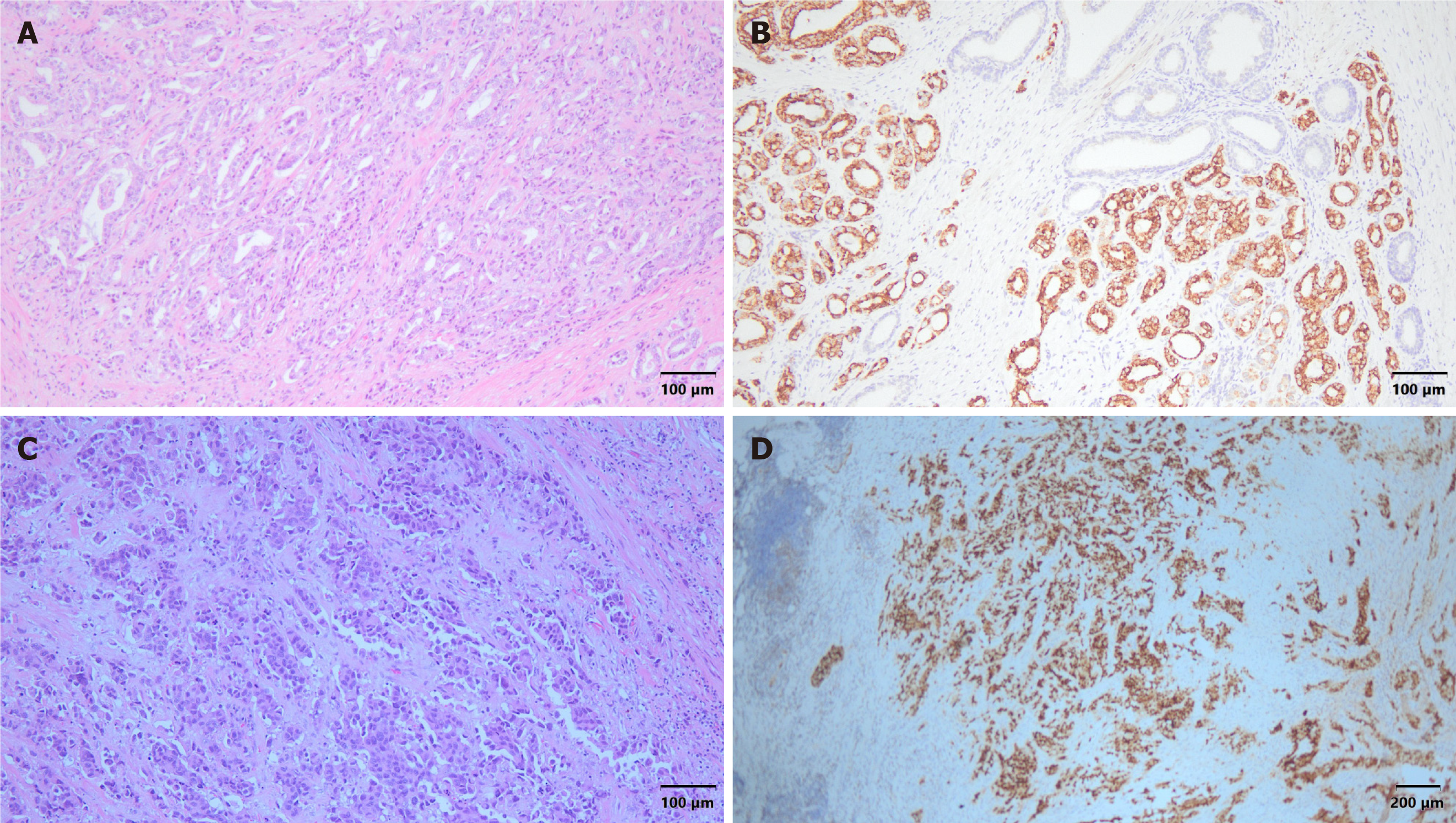
Figure 6 Histopathological analysis and immunohistochemical examination of the resected specimen.
A: Adenocarcinoma of the prostate (hematoxylin and eosin staining × 100); B: Positive immunohistochemical (IHC) staining for α-methylacyl coenzyme A racemase ( × 200); C: Prostate urothelial carcinoma (hematoxylin and eosin staining × 100); D: Positive IHC staining for GATA binding protein 3 ( × 40).
- Citation: Hsu JY, Lin YS, Huang LH, Tsao TY, Hsu CY, Ou YC, Tung MC. Concurrent occurrence of adenocarcinoma and urothelial carcinoma of the prostate gland: A case report. World J Clin Cases 2024; 12(26): 5952-5959
- URL: https://www.wjgnet.com/2307-8960/full/v12/i26/5952.htm
- DOI: https://dx.doi.org/10.12998/wjcc.v12.i26.5952


