Copyright
©The Author(s) 2024.
World J Clin Cases. Sep 16, 2024; 12(26): 5946-5951
Published online Sep 16, 2024. doi: 10.12998/wjcc.v12.i26.5946
Published online Sep 16, 2024. doi: 10.12998/wjcc.v12.i26.5946
Figure 1
Periorbital purpura was visible.
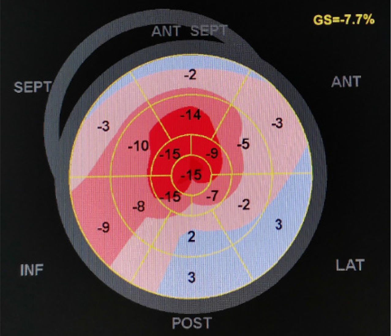
Figure 2 Two-dimensional speckle tracking strain and strain rate imaging of the myocardium: Visible apical exemption phenomenon is visible.
ANT: Anterior; GS: Global strain; INF: Inferior; LAT: Anterolateral; POST: Inferolateral; SEPT: Posterior septum.
Figure 3
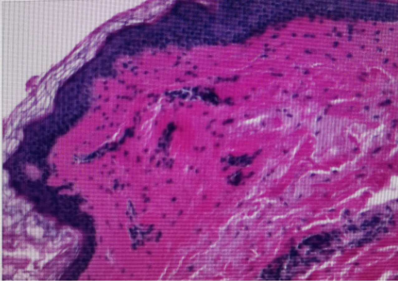
Amyloid deposition in collagen fibers of the dermis and positive Congo red staining.
- Citation: Wang XF, Li T, Yang M, Huang Y. Periorbital purpura can be the only initial symptom of primary light chain amyloidosis: A case report. World J Clin Cases 2024; 12(26): 5946-5951
- URL: https://www.wjgnet.com/2307-8960/full/v12/i26/5946.htm
- DOI: https://dx.doi.org/10.12998/wjcc.v12.i26.5946


