Copyright
©The Author(s) 2024.
World J Clin Cases. Sep 6, 2024; 12(25): 5784-5790
Published online Sep 6, 2024. doi: 10.12998/wjcc.v12.i25.5784
Published online Sep 6, 2024. doi: 10.12998/wjcc.v12.i25.5784
Figure 1 Pathological analysis of a sinonasal teratocarcinosarcoma.
A: Low magnification of hematoxylin-eosin stain showing the overall variegated appearance (40 ×); B: Glandular epithelial structures were seen within the tumor as well as focal nested squamous epithelial components, with translucent cytoplasm and no keratinization (100 ×); C: Localized patchy glandular epithelium structures were seen in localized lesion location (100 ×); D: Under high magnification, tumor cells exhibited significant heterogeneity, with readily observable nuclear division figures (400 ×).
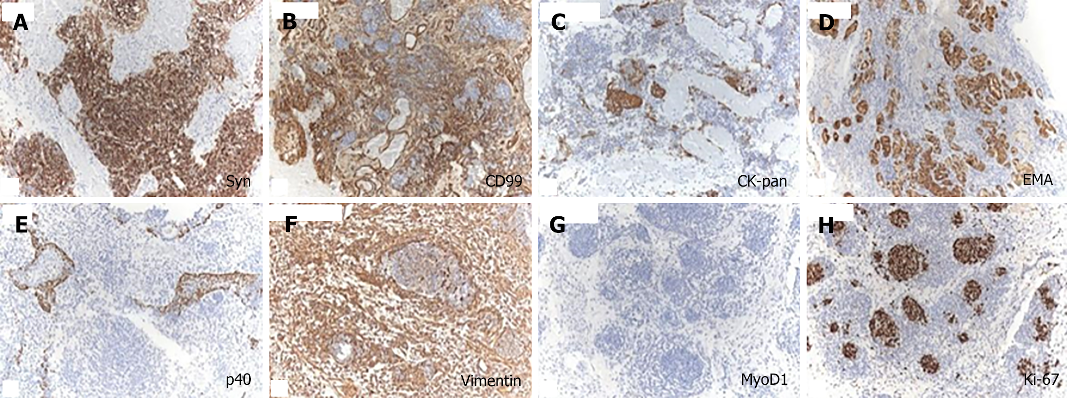
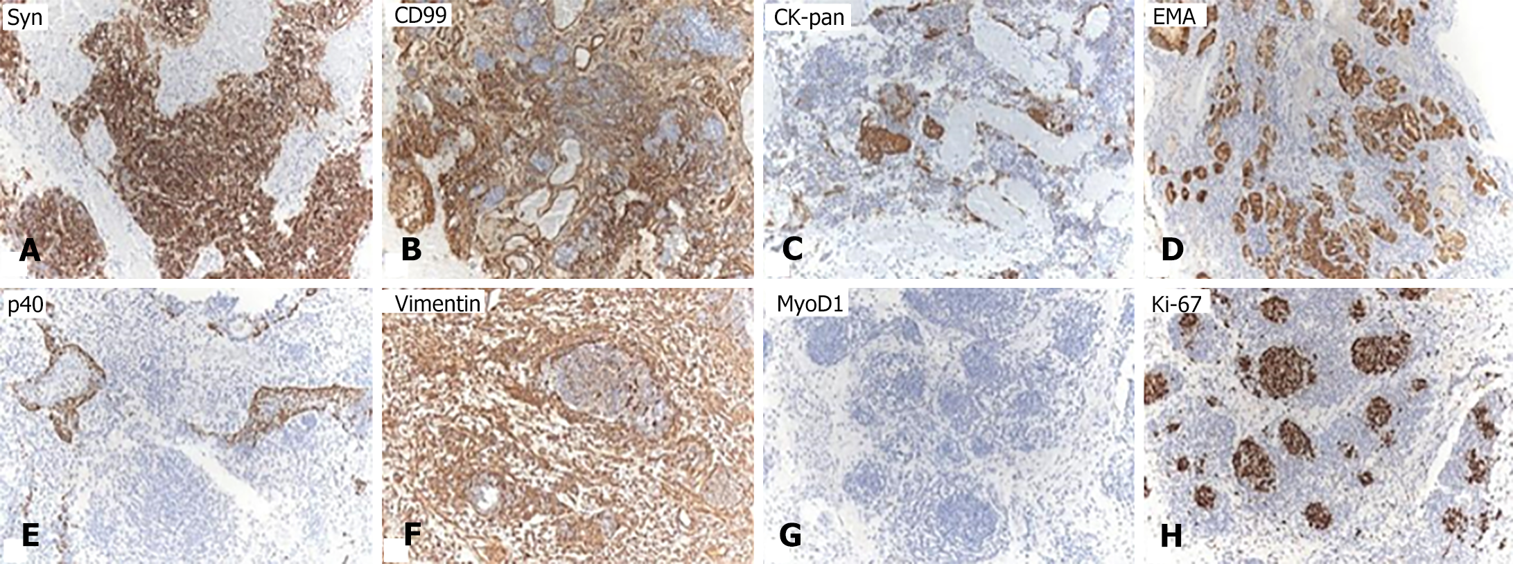
Figure 2 The tumor and neural epithelia were negative.
A: Syn; B: CD99; C: CK-pan; D: Epithelial membrane antigen; E: P40; F: Vimentin; G: MyoD1; H: 40% positive for Ki-67.
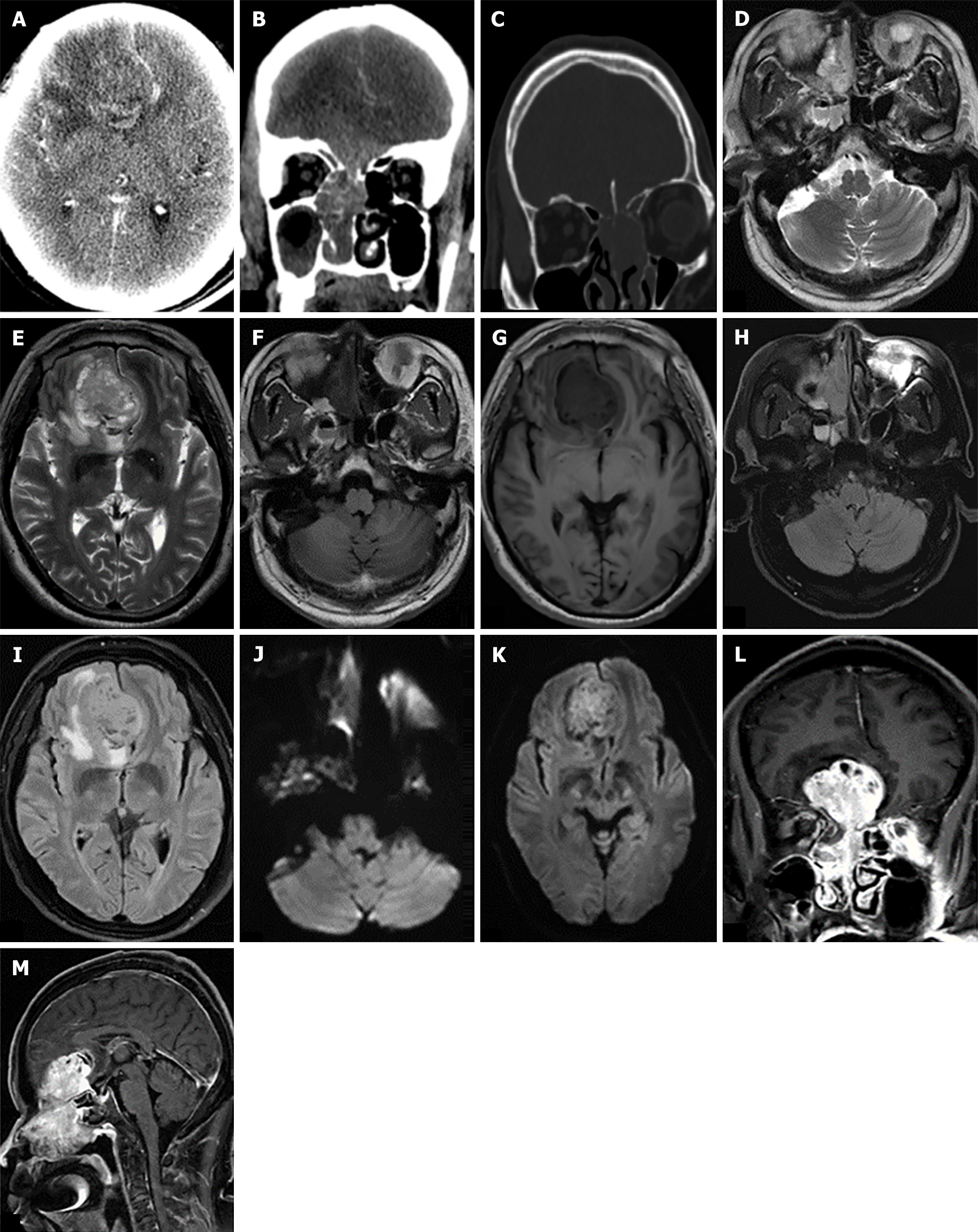
Figure 3 Computed tomography and magnetic resonance imaging scan images.
A: Axial enhanced computed tomography (CT) scan revealed an irregularly enhanced tumor resembling a mass in the right frontal region; B: Coronal enhanced CT scan revealed a polypoid mass with expansive growth in the right nasal cavity, extending intracranially across the cribriform plate; C: Sagittal CT bone window revealed multiple bone destruction in bilateral skull base and ethmoid sinus wall; D-K: Magnetic resonance imaging (MRI) scan reveals a mass-like abnormal signal in the right nasal cavity and right frontal region, characterized by slightly increased signal intensity on T2-weighted images, slightly decreased signal intensity on T1-weighted images, unevenly slightly increased signal intensity on FLAIR images, and slightly increased signal intensity on diffusion-weighted imaging. There is hyperintensity in the frontal lobe adjacent to tumor (vasogenic edema/infiltration); L and M: The enhanced MRI scan revealed a diffusely heterogeneous mass with enhanced contrast in the right nasal cavity, infiltrating into the cranial cavity beyond the cribriform plate.
- Citation: Fu LY, Yang MY, Ye PY, Wang ZC, Chen CJ, Li H, Xu SW. Sinonasal teratocarcinosarcoma involving the nasal cavity, unilateral paranasal sinuses, and intracranial invasion: A case report. World J Clin Cases 2024; 12(25): 5784-5790
- URL: https://www.wjgnet.com/2307-8960/full/v12/i25/5784.htm
- DOI: https://dx.doi.org/10.12998/wjcc.v12.i25.5784


