Copyright
©The Author(s) 2024.
World J Clin Cases. Aug 26, 2024; 12(24): 5613-5621
Published online Aug 26, 2024. doi: 10.12998/wjcc.v12.i24.5613
Published online Aug 26, 2024. doi: 10.12998/wjcc.v12.i24.5613
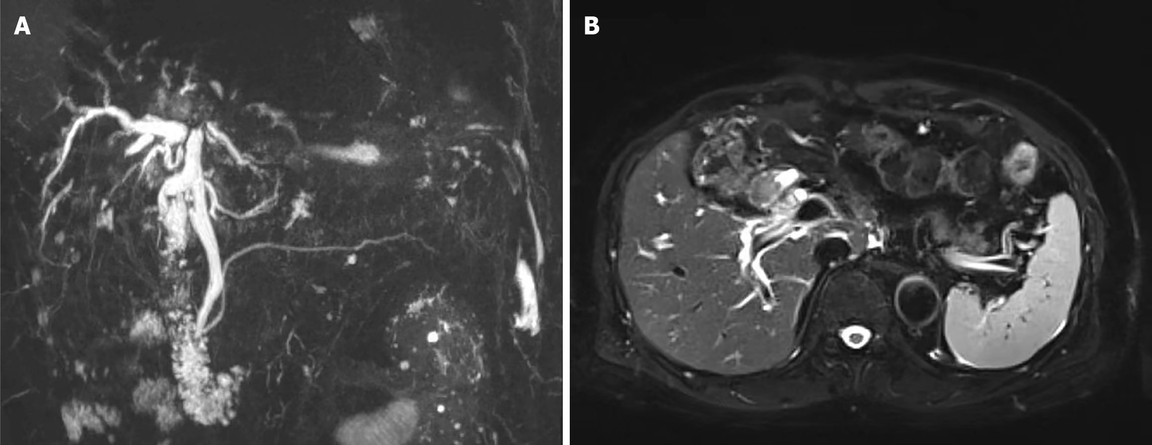
Figure 1 Admission magnetic resonance cholangiopancreatography and magnetic resonance imaging.
A: Magnetic resonance cholangiopan
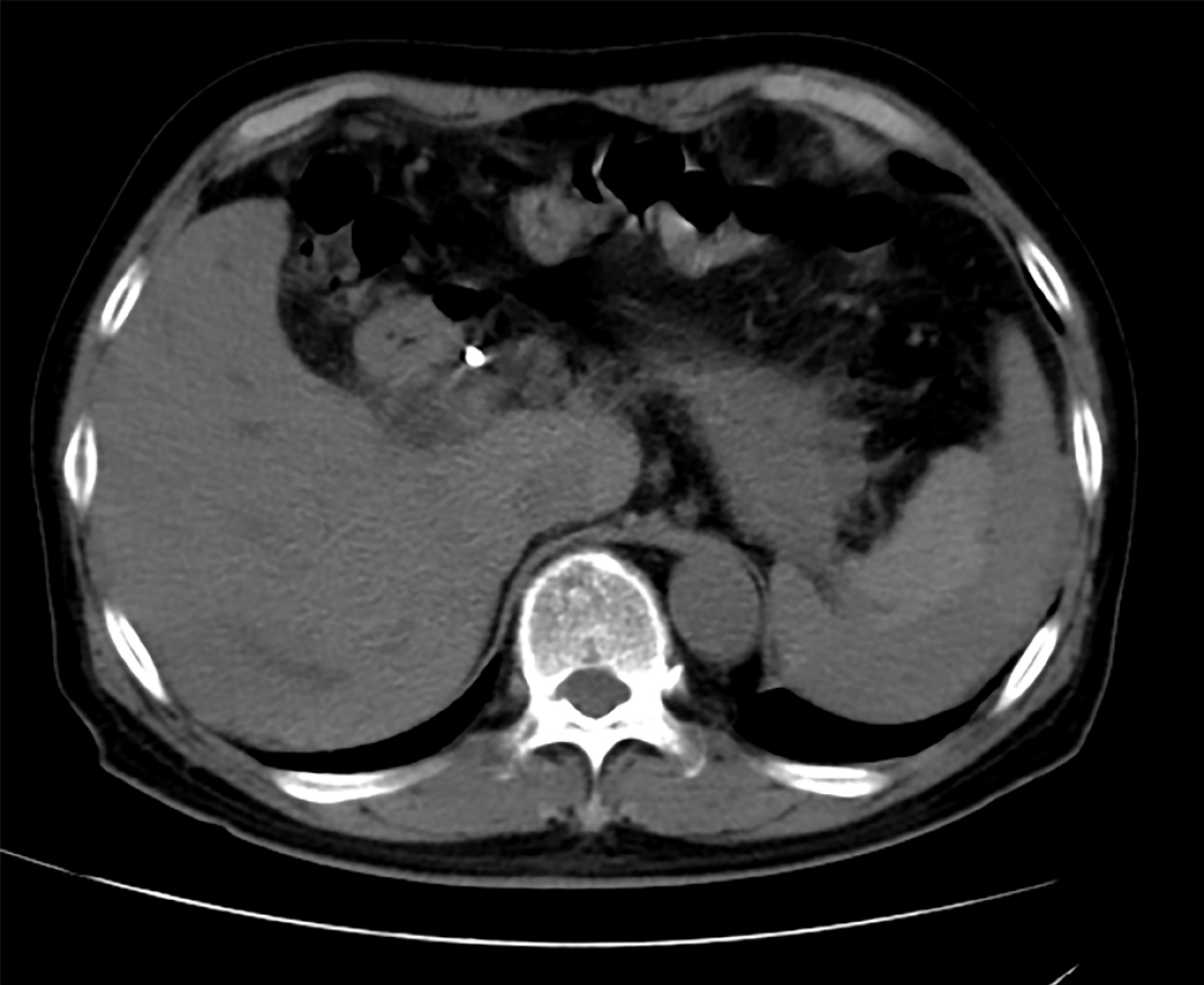
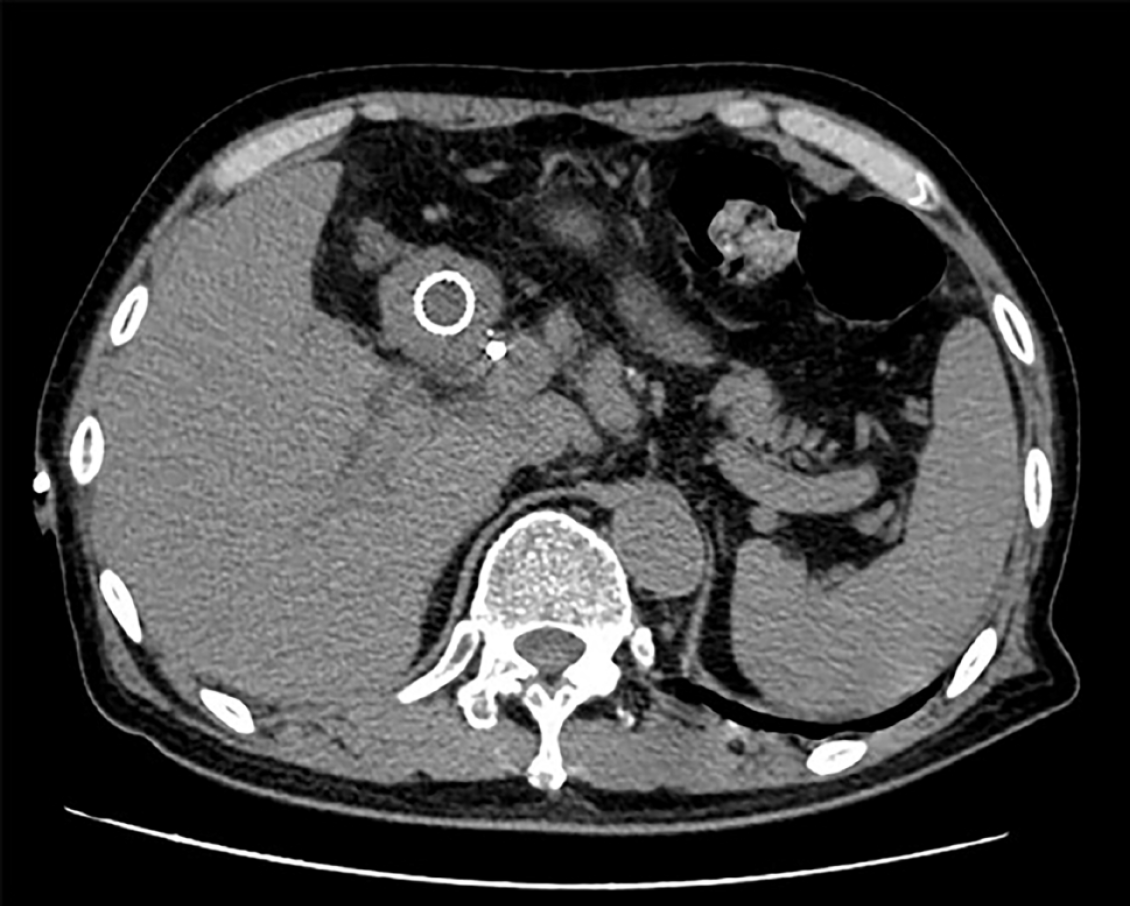
Figure 2
Computed tomography scan revealed a hematoma in the spleen after endoscopic retrograde cholangiopancreatography.
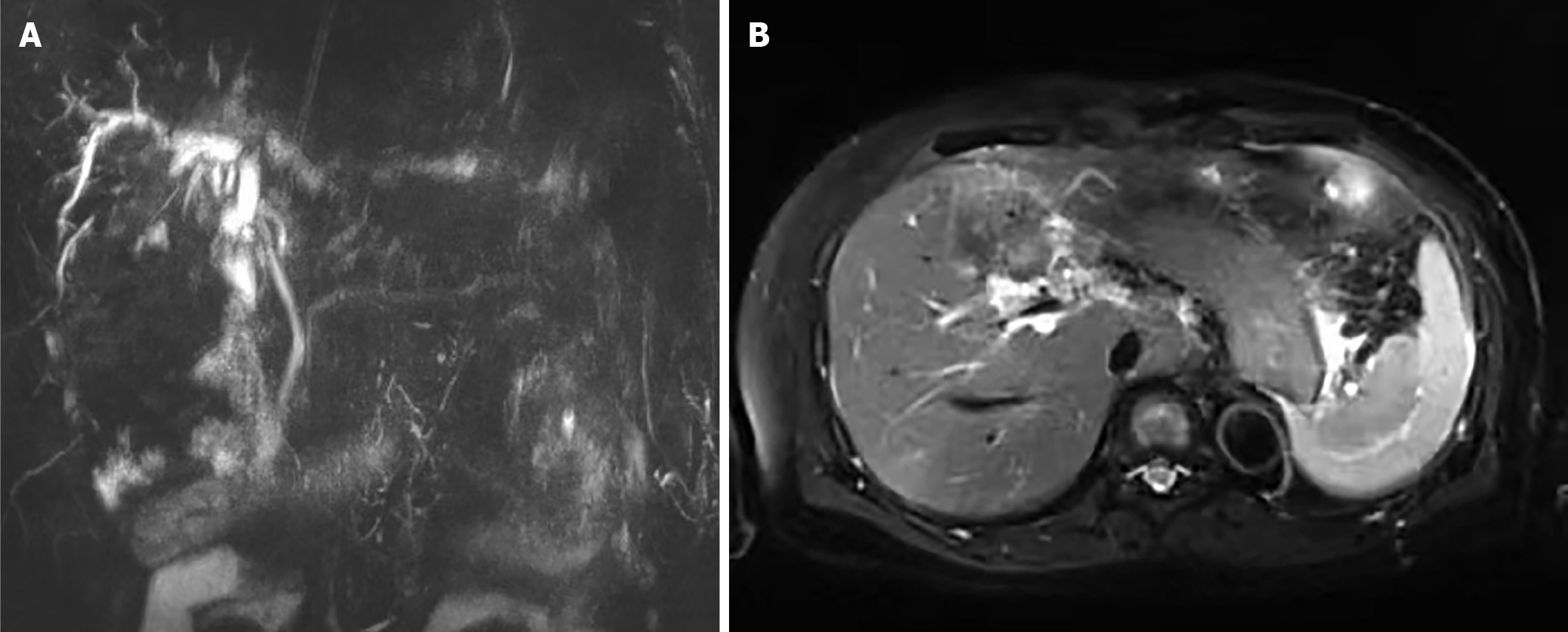
Figure 3 Magnetic resonance cholangiopancreatography and magnetic resonance imaging after endoscopic retrograde cholangiopan
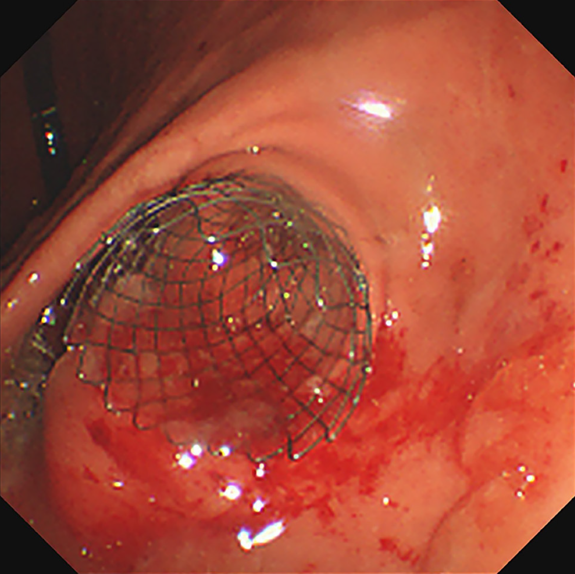
Figure 4
Endoscopic pyloric stent placement with minor intraoperative bleeding.
Figure 5
A computed tomography scan conducted before discharge showed that the splenic hematoma had disappeared.
- Citation: Guo CY, Wei YX. Splenic subcapsular hematoma following endoscopic retrograde cholangiopancreatography: A case report and review of literature. World J Clin Cases 2024; 12(24): 5613-5621
- URL: https://www.wjgnet.com/2307-8960/full/v12/i24/5613.htm
- DOI: https://dx.doi.org/10.12998/wjcc.v12.i24.5613


