Copyright
©The Author(s) 2024.
World J Clin Cases. Aug 16, 2024; 12(23): 5416-5421
Published online Aug 16, 2024. doi: 10.12998/wjcc.v12.i23.5416
Published online Aug 16, 2024. doi: 10.12998/wjcc.v12.i23.5416
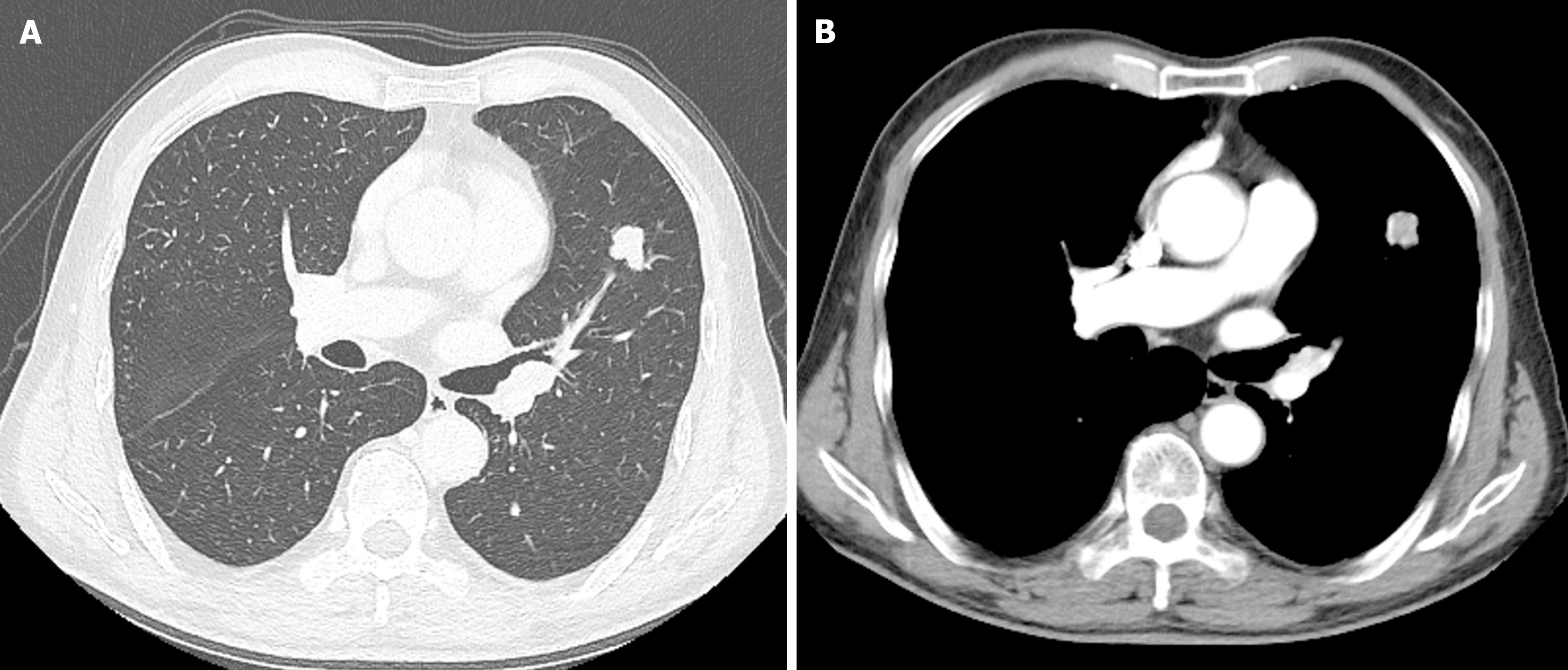
Figure 1 A chest enhanced computed tomography scan revealing a soft tissue nodule in the upper lobe of the left lung.
A: Lung window; B: Mediastinal window.
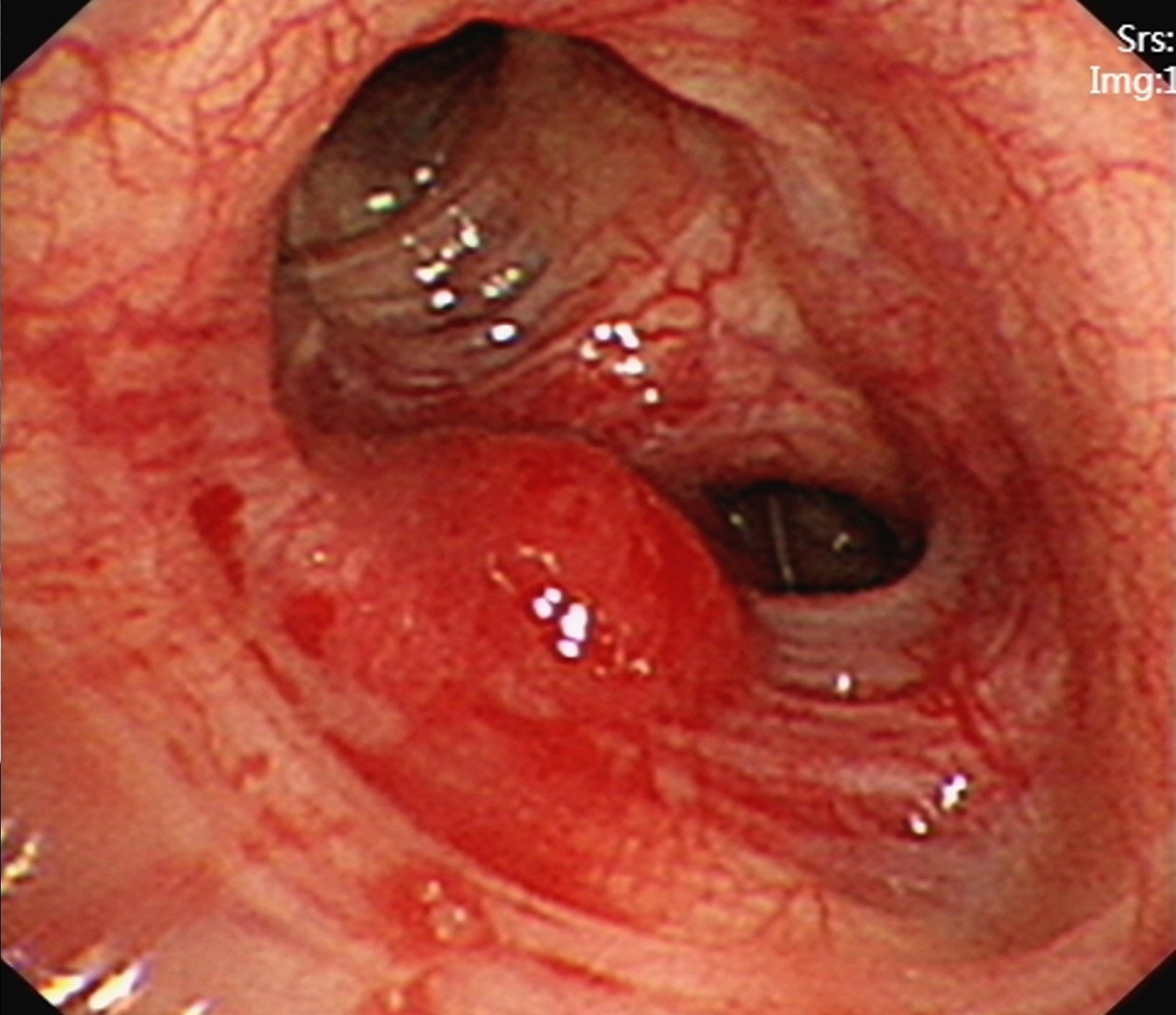
Figure 2
Flexible bronchoscopy revealing a hypervascular lesion in the bronchus of the left superior lobe of the lung.
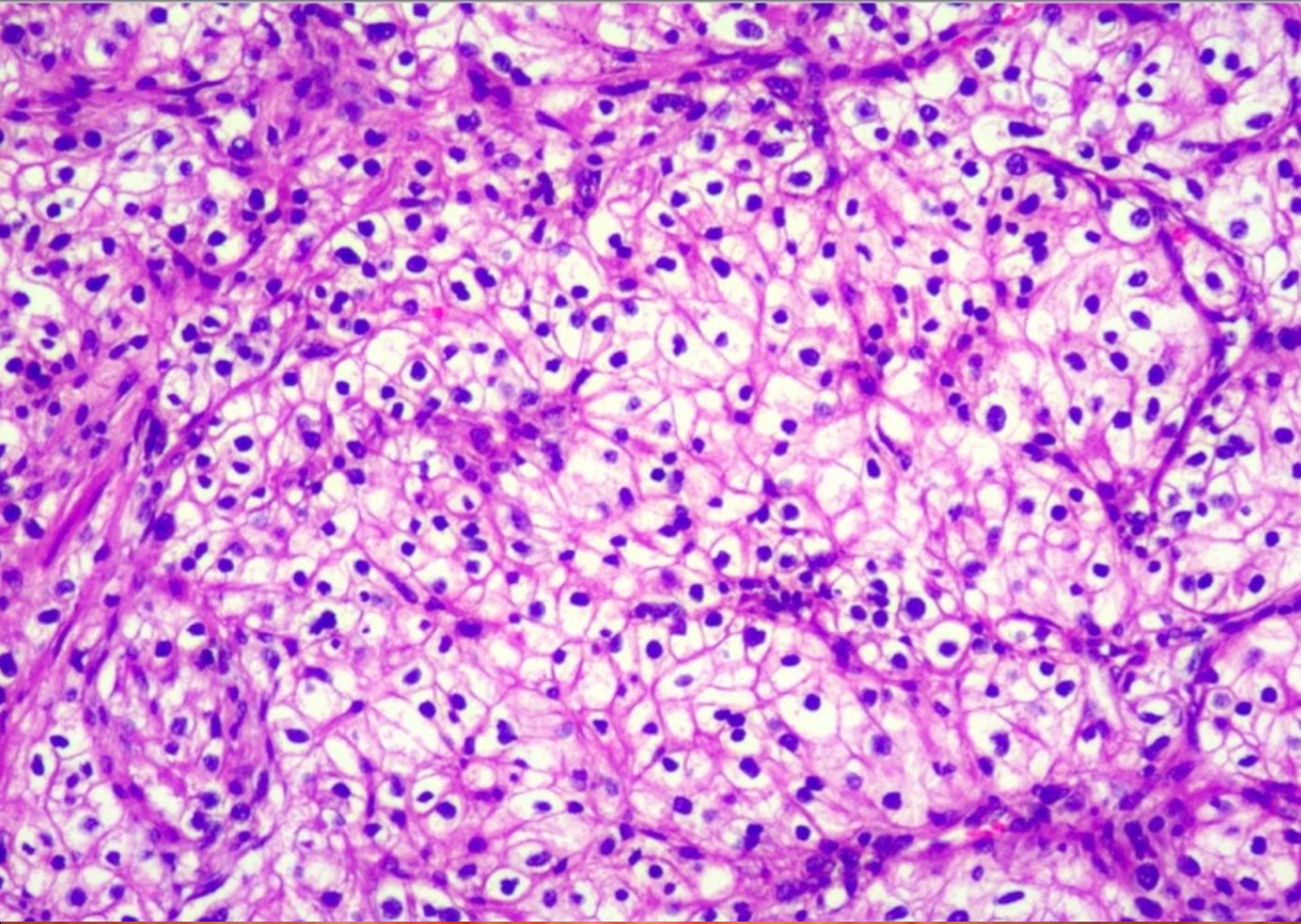
Figure 3
Haematoxylin and eosin-stained section of the nodule biopsy showing renal clear cell carcinoma.
- Citation: Xie TH, Fu Y, Ha SN, Meng QX, Sun Q, Wang P. Endobronchial metastasis secondary to renal clear cell carcinoma: A case report. World J Clin Cases 2024; 12(23): 5416-5421
- URL: https://www.wjgnet.com/2307-8960/full/v12/i23/5416.htm
- DOI: https://dx.doi.org/10.12998/wjcc.v12.i23.5416


