Copyright
©The Author(s) 2024.
World J Clin Cases. Aug 6, 2024; 12(22): 5276-5282
Published online Aug 6, 2024. doi: 10.12998/wjcc.v12.i22.5276
Published online Aug 6, 2024. doi: 10.12998/wjcc.v12.i22.5276
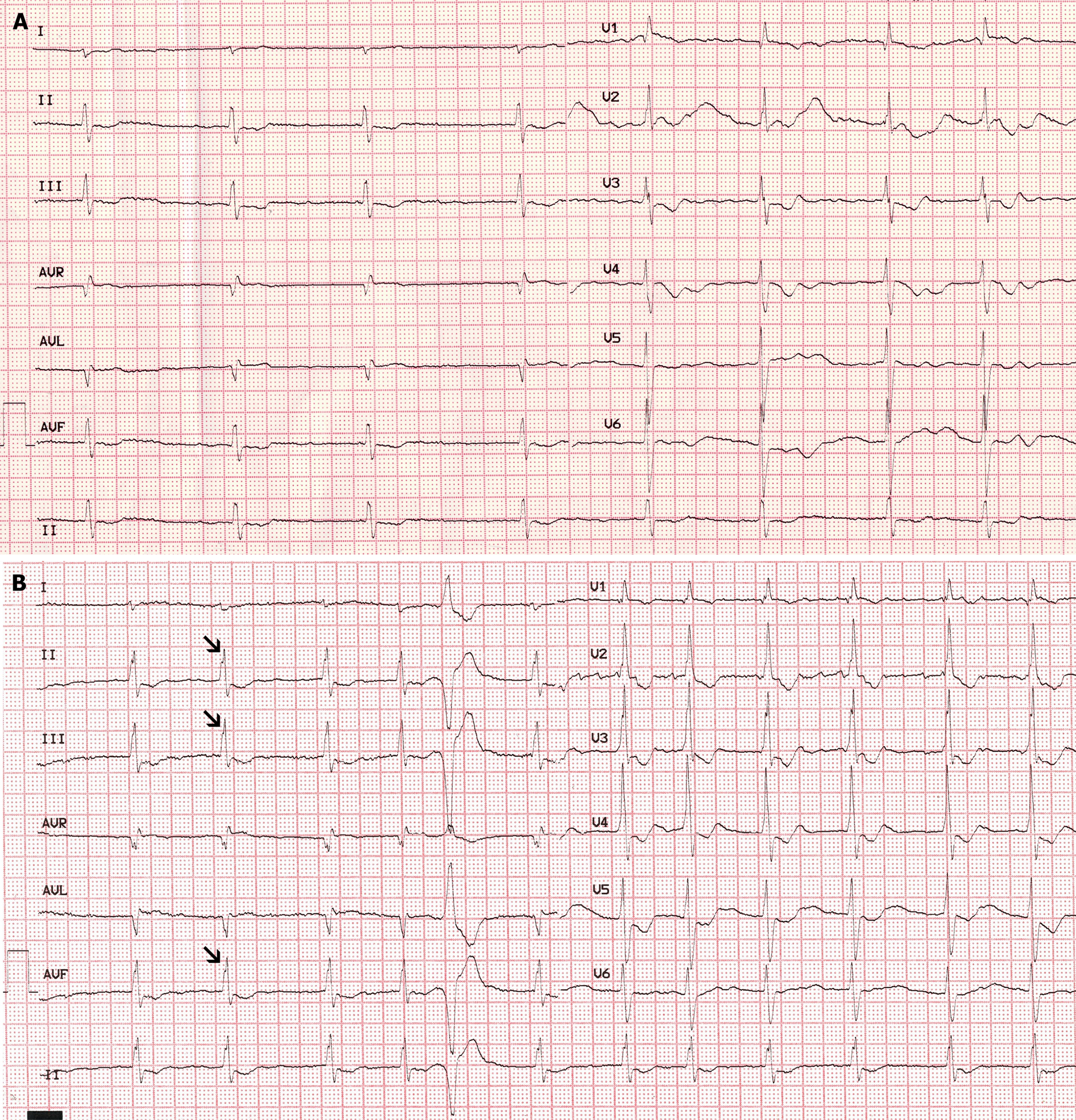
Figure 1 Initial electrocardiogram.
A: Atrial fibrillation with a long relative risk interval; B: Atrial fibrillation with crochetage pattern in leads II, III, and aVF and incomplete right bundle branch block (arrows pointing toward crochetage sign are most common).
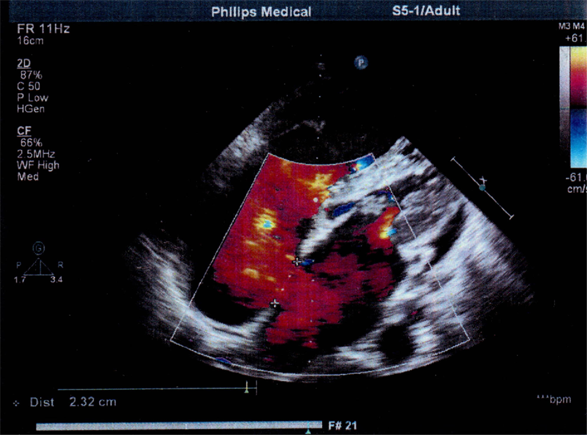
Figure 2 Echocardiographic image.
Echocardiography shows a secundum atrial septal defect that measured 2.32 cm.
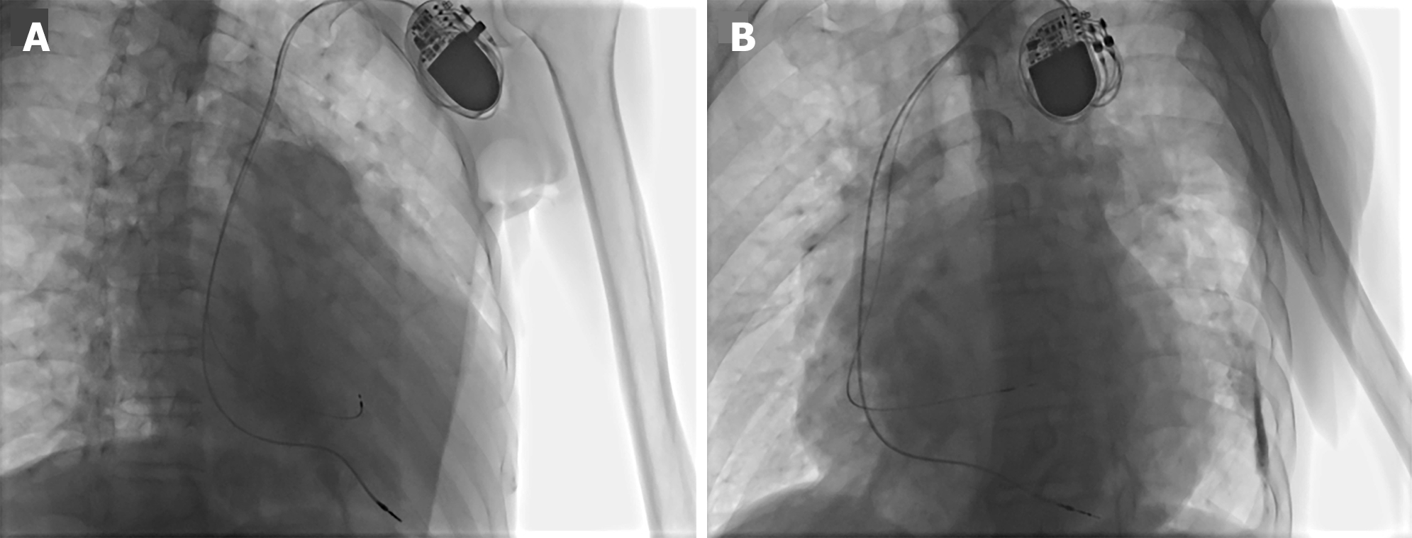
Figure 3 Fluoroscopic image.
The fluoroscopic image shows that the 3830 lead was located in the His-bundle region. A: Right-anterior-oblique 30° view; B: Left-anterior-oblique 40° view.
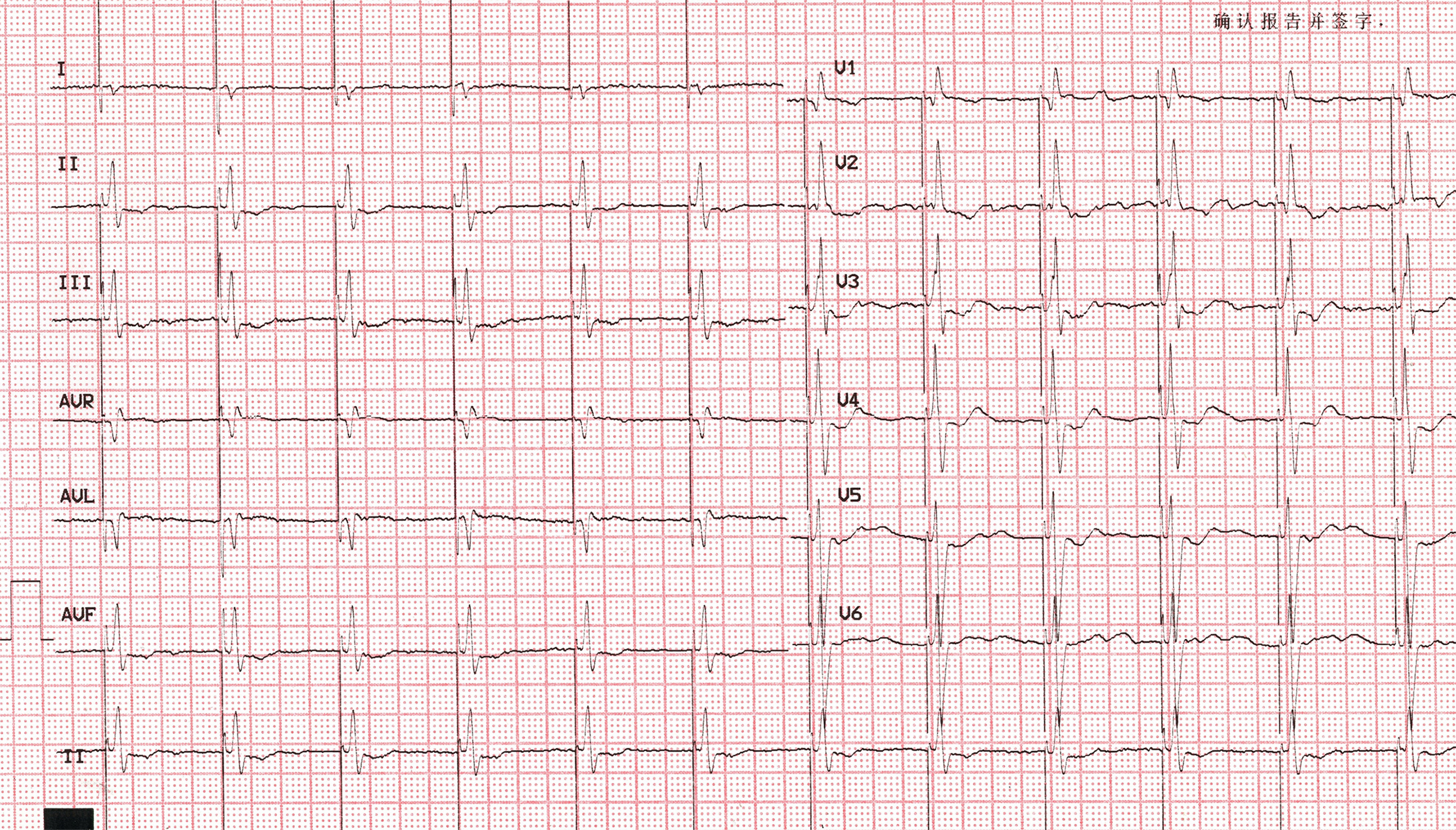
Figure 4 Postoperative electrocardiogram.
Electrocardiogram showing the disappearance of crochetage sign after his-bundle pacing.
- Citation: Mu YG, Liu KS. Selective his bundle pacing eliminates crochetage sign: A case report. World J Clin Cases 2024; 12(22): 5276-5282
- URL: https://www.wjgnet.com/2307-8960/full/v12/i22/5276.htm
- DOI: https://dx.doi.org/10.12998/wjcc.v12.i22.5276


