Copyright
©The Author(s) 2024.
World J Clin Cases. Aug 6, 2024; 12(22): 5189-5195
Published online Aug 6, 2024. doi: 10.12998/wjcc.v12.i22.5189
Published online Aug 6, 2024. doi: 10.12998/wjcc.v12.i22.5189
Figure 1 Intraoral examination photos of the case.
A: Full-mouth view; B: Maxillary arch view; C: Mandibular arch view.
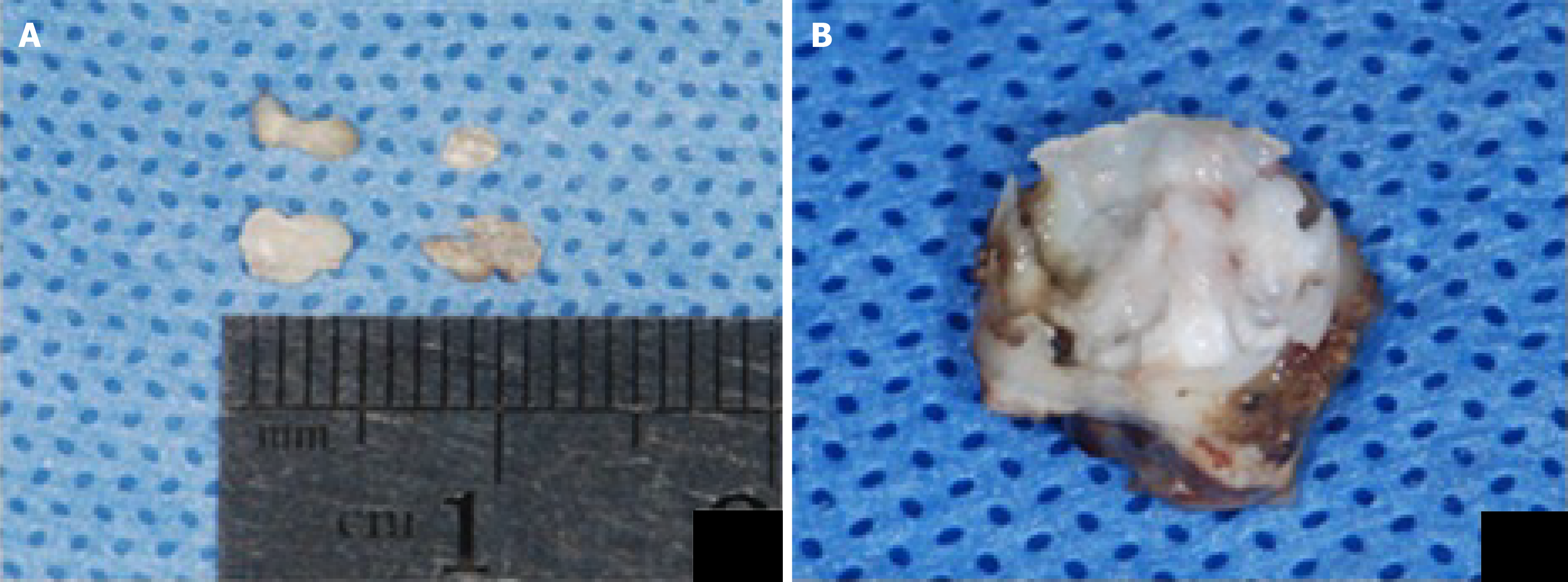
Figure 2 Pulp stone findings and treatments.
A: Pulp stones extracted from tooth 46 during the initial visit, showing partial removal; B: Irregular calcifications observed in the pulp cavity of the extracted tooth 85 during the follow-up visit in November 2022, after completing root canal treatment for tooth 46.
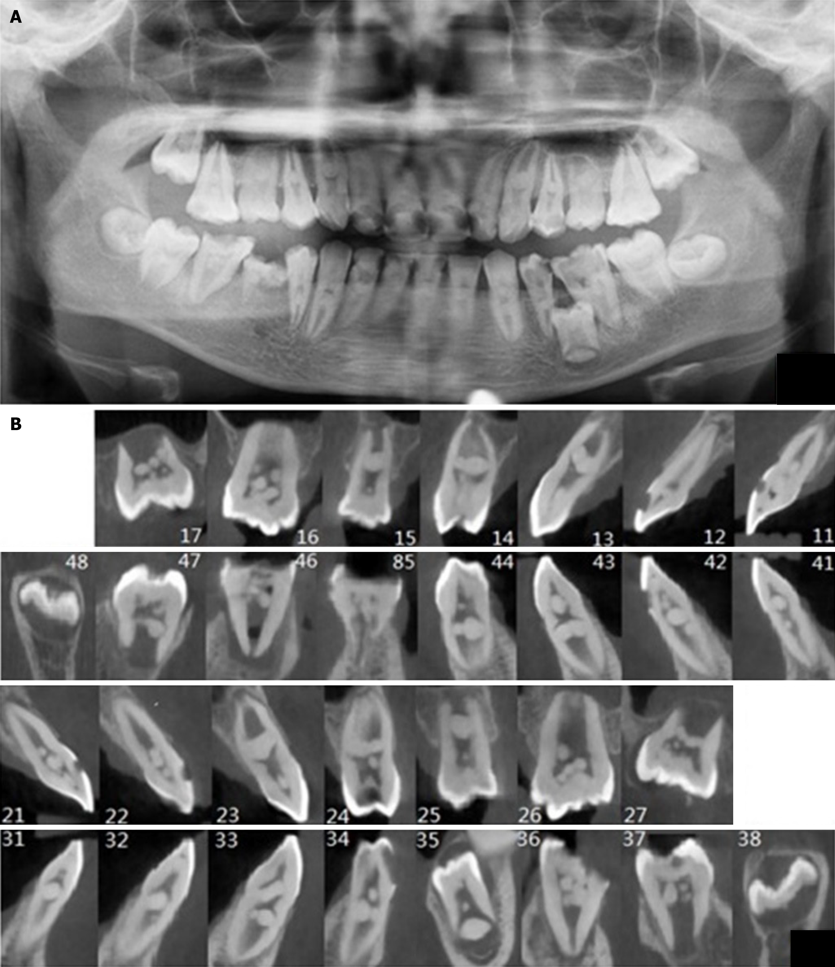
Figure 3 Radiographic images of the case during the initial visit in October 2020.
A: Panoramic radiograph; B: Cone-beam computed tomography scan of the teeth.
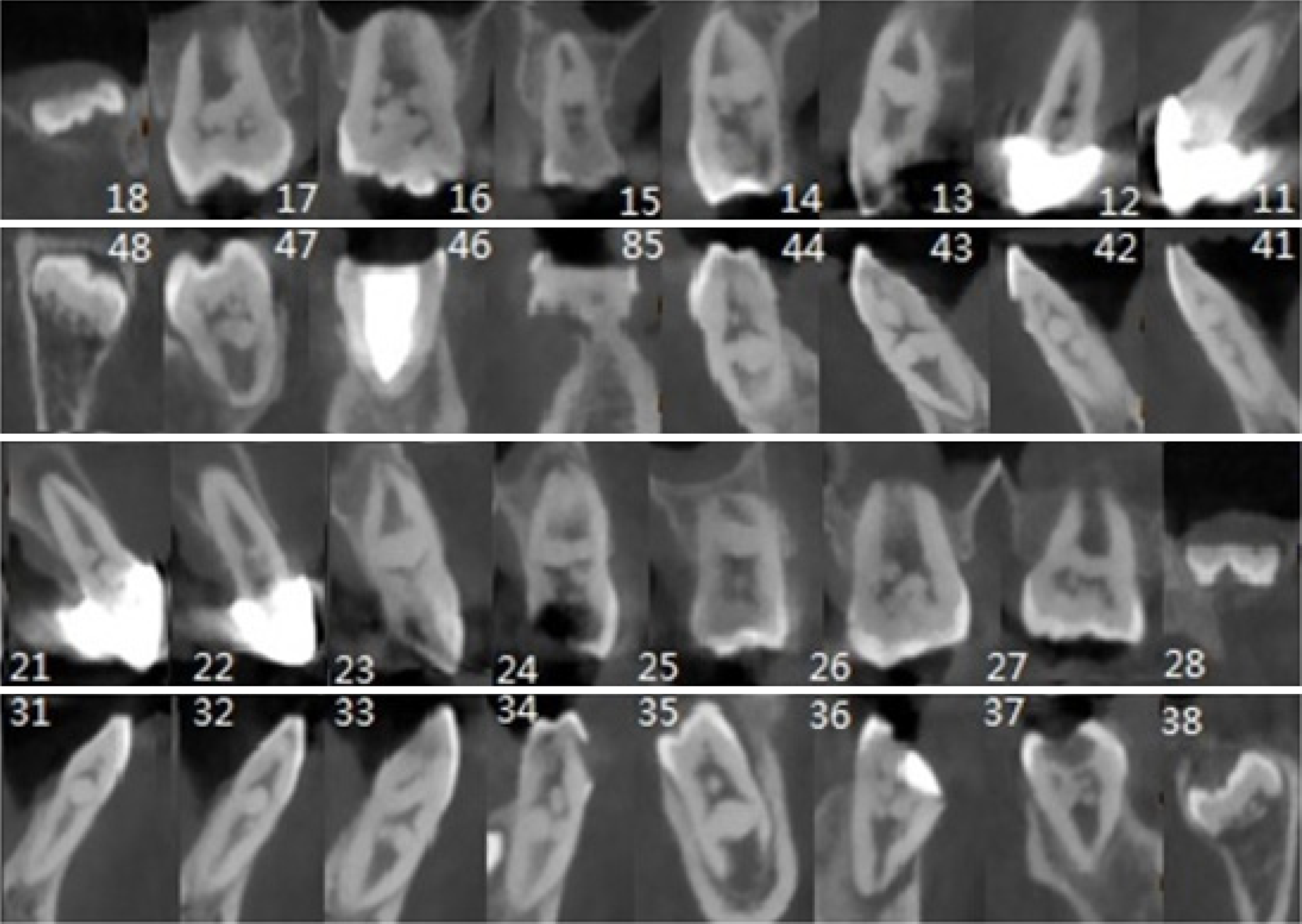
Figure 4 Cone-beam Computed Tomography scan of all teeth (In December 2022).
- Citation: Lv Y, Zhu J, Fu CT, Liu L, Wang J, Li YF. Multiple pulp stones emerge across all teeth during mixed dentition: A case report. World J Clin Cases 2024; 12(22): 5189-5195
- URL: https://www.wjgnet.com/2307-8960/full/v12/i22/5189.htm
- DOI: https://dx.doi.org/10.12998/wjcc.v12.i22.5189


