Copyright
©The Author(s) 2024.
World J Clin Cases. Aug 6, 2024; 12(22): 5168-5176
Published online Aug 6, 2024. doi: 10.12998/wjcc.v12.i22.5168
Published online Aug 6, 2024. doi: 10.12998/wjcc.v12.i22.5168
Figure 1 Ultrasound findings.
A: Right ovarian echoless area with clear boundary; B: No color blood flow signal was found in the sac wall of the right ovarian anechoic region; C: Solid mass in the left adnexal area with clear boundary; D: Abundant color blood flow signals were seen around the solid mass in the left adnexal area.
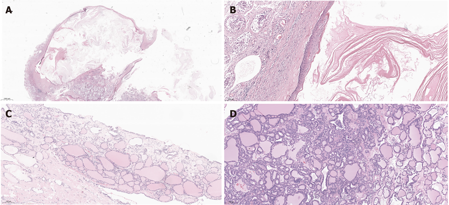
Figure 2 Pathological findings.
A: Histological section of the epidermoid cyst of the right testicle showing the structure of the cyst (hematoxylin & eosin × 1); B: The cyst wall is composed of squamous epithelium surrounding the lamellar keratin layer, and there is a large amount of keratin in the cyst cavity (hematoxylin & eosin × 10); C: Histological section of the right ovarian cyst showed a large amount of thyroid follicular tissue (hematoxylin & eosin × 10); D: Histological sections of the left ovary showed a large amount of thyroid follicular tissue (hematoxylin & eosin × 10)
- Citation: He LY, Li W. Monodermal teratoma: Three case reports and review of literature. World J Clin Cases 2024; 12(22): 5168-5176
- URL: https://www.wjgnet.com/2307-8960/full/v12/i22/5168.htm
- DOI: https://dx.doi.org/10.12998/wjcc.v12.i22.5168


