Copyright
©The Author(s) 2024.
World J Clin Cases. Aug 6, 2024; 12(22): 5145-5150
Published online Aug 6, 2024. doi: 10.12998/wjcc.v12.i22.5145
Published online Aug 6, 2024. doi: 10.12998/wjcc.v12.i22.5145
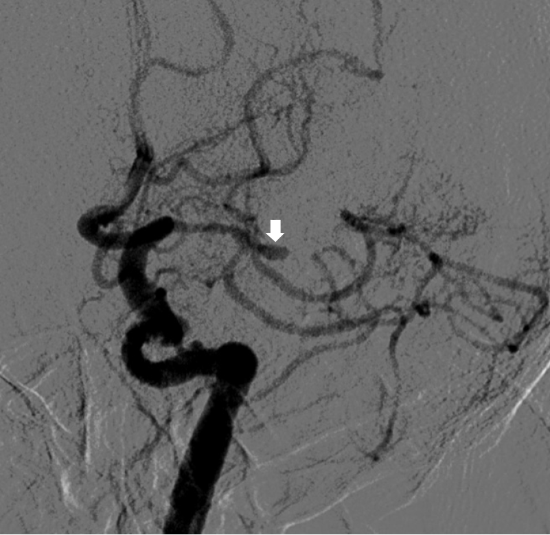
Figure 1 Preoperative digital subtraction cerebral angiography image of the aneurysm.
White arrow: Arterial aneurysm at the bifurcation of the left middle cerebral artery.
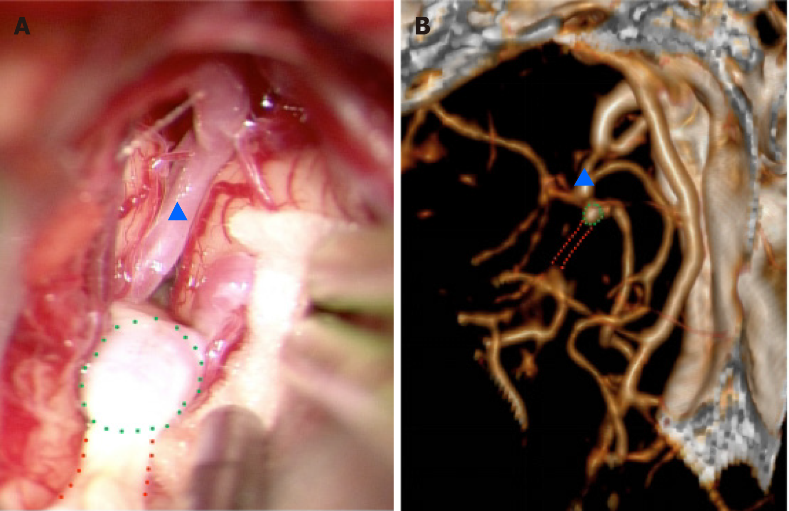
Figure 2 Intraoperative photograph and preoperative computed tomography angiography image.
A: A photograph captured under a microscope during surgery; B: A computed tomography angiography image of the same viewing angle. The green dots outline the arterial stump caused by occlusion of the middle cerebral artery, which has a sac-like, dilated appearance resembling an aneurysm; the red dots outline the distal occluded part of the middle cerebral artery, which extends to the lateral fissure and anastomoses with other arteries. The arrowhead indicates the narrowed M1 segment of the middle cerebral artery.
Figure 3 Preoperative digital subtraction cerebral angiography image of the collateral circulation.
A: The enlarged remnant of the left middle cerebral artery and the distal vessels form an anastomosis in the lateral fissure; B and C: The region was supplied by the cortical leptomeningeal branch of the anterior cerebral artery, which was previously the territory of the middle cerebral artery.
- Citation: Yang S, Mai RK. Mimicking aneurysm in a patient with chronic occlusion of the left middle cerebral artery: A case report. World J Clin Cases 2024; 12(22): 5145-5150
- URL: https://www.wjgnet.com/2307-8960/full/v12/i22/5145.htm
- DOI: https://dx.doi.org/10.12998/wjcc.v12.i22.5145


