Copyright
©The Author(s) 2024.
World J Clin Cases. Aug 6, 2024; 12(22): 4932-4939
Published online Aug 6, 2024. doi: 10.12998/wjcc.v12.i22.4932
Published online Aug 6, 2024. doi: 10.12998/wjcc.v12.i22.4932
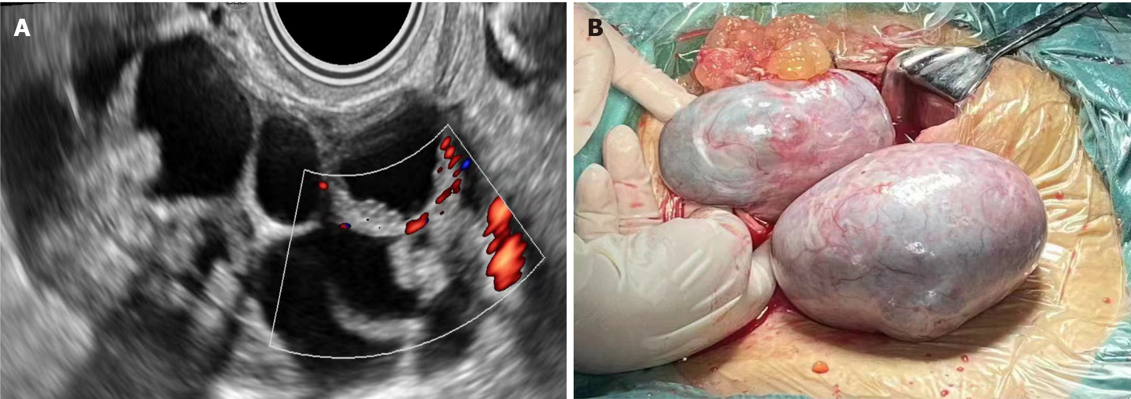
Figure 1 Ultrasound images and surgical images of borderline or focal borderline ovarian collision tumors.
A: Bilateral ovarian collision tumor, serous cystadenoma with fibroma, vegetable pattern can be seen outside the left cyst wall (classified as type 3 ultrasound phenotype); B: Direct view of serous cystadenoma with fibroma surgery.
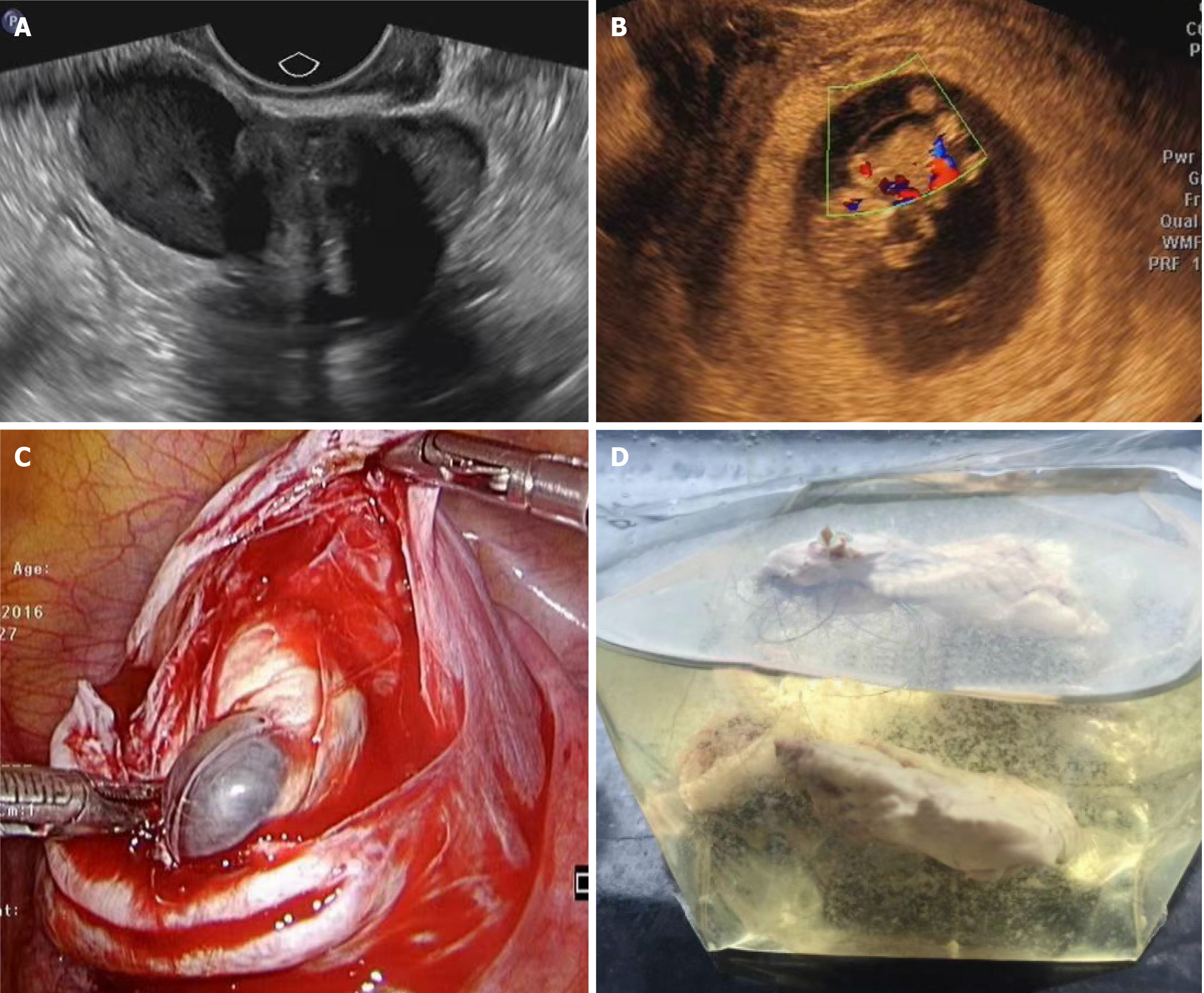
Figure 2 Ultrasound image and surgical image of ovarian collision tumor.
A: Endometriotic cyst with fibroma, consisting of two typical sonographic phenotypic features, collision tumor (classified as class 1 sonographic phenotype); B: The large cyst in the cyst is serous cystadenoma, and the small inner cyst is goiter (classified as type 2 ultrasound phenotype); C: Intraoperative findings of cyst sac: A 4-cm small cyst can be seen in the denuded large cyst; D: Gross specimen of cyst capsule, with serous cystadenoma cyst wall at the bottom and teratoma hair at the upper layer.
- Citation: Yin C, Wang Y, Fei ZH, Sun LH, Zhou WA, Li H. Ovarian-adnexal reporting and data system ultrasound evaluation and pathological characteristics of ovarian collision tumor. World J Clin Cases 2024; 12(22): 4932-4939
- URL: https://www.wjgnet.com/2307-8960/full/v12/i22/4932.htm
- DOI: https://dx.doi.org/10.12998/wjcc.v12.i22.4932


