Copyright
©The Author(s) 2024.
World J Clin Cases. Jul 26, 2024; 12(21): 4807-4812
Published online Jul 26, 2024. doi: 10.12998/wjcc.v12.i21.4807
Published online Jul 26, 2024. doi: 10.12998/wjcc.v12.i21.4807
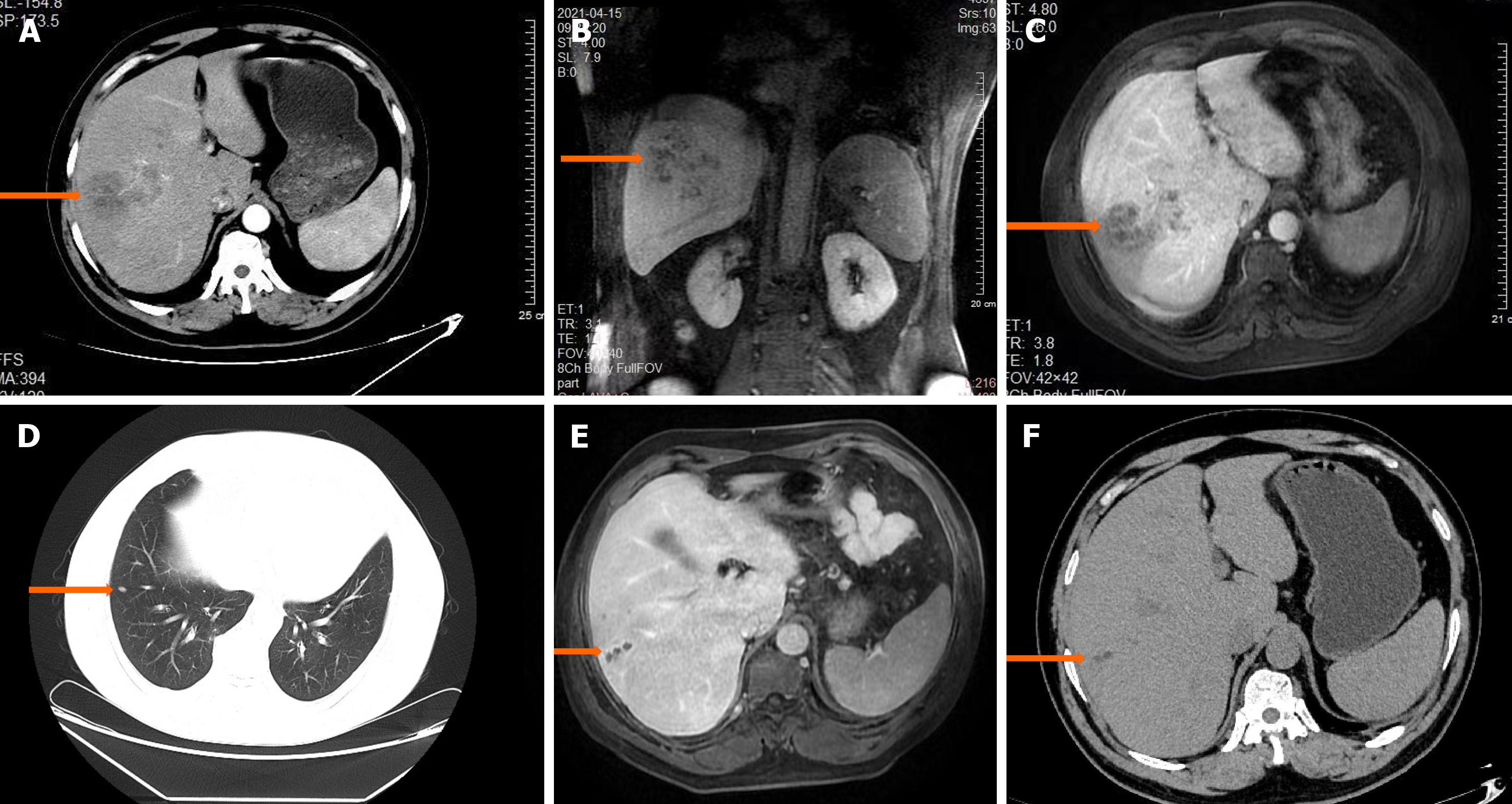
Figure 1 Abdominal enhanced computed tomography and magnetic resonance imaging.
A: In April 2021, abdominal enhanced computed tomography (CT) of the right lobe of the liver showed a mass of low density; B: In April 2021, magnetic resonance imaging (MRI) in the coronal position showed an uneven flaky shadow in the liver parenchyma; C: In April 2021, MRI T1-weighted imaging (T1WI) showed that the lesion in the right lobe of the liver was mildly intensified; D: In November 2021, chest CT showed small nodular foci in the middle lobe of the right lung; E: In November 2021, abdominal MRI showed a bunch of grapes sign in T1WI in the right lobe of the liver; F: In March 2022, abdominal CT indicated that the lesion was smaller than before.
- Citation: Zheng YQ, Guo GB, Wang MF, Zhu HZ, Zhou C, Li LH, Zhang L, Liu YQ. Paragonimiasis misdiagnosed as liver abscess: A case report. World J Clin Cases 2024; 12(21): 4807-4812
- URL: https://www.wjgnet.com/2307-8960/full/v12/i21/4807.htm
- DOI: https://dx.doi.org/10.12998/wjcc.v12.i21.4807


