Copyright
©The Author(s) 2024.
World J Clin Cases. Jul 26, 2024; 12(21): 4794-4801
Published online Jul 26, 2024. doi: 10.12998/wjcc.v12.i21.4794
Published online Jul 26, 2024. doi: 10.12998/wjcc.v12.i21.4794
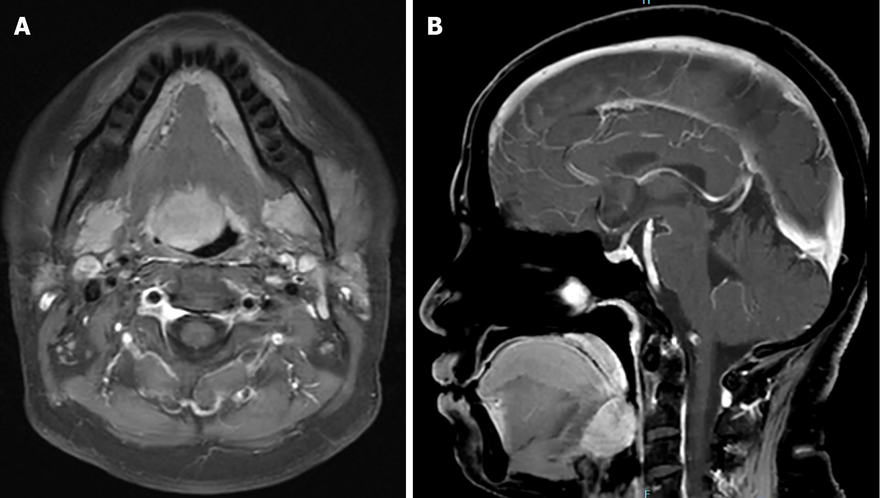
Figure 1 Magnetic resonance imaging showing a mass at the base of the tongue with marked homogeneous enhancement.
A: Axial image showed a mass located to the right of the root of the tongue; B: Sagittal image showed a mass at the base of the tongue.
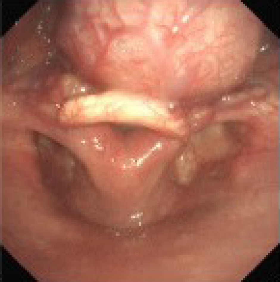
Figure 2
Laryngoscopy showing a mass at the base of the tongue, above the epiglottis, with smooth surface mucosa.
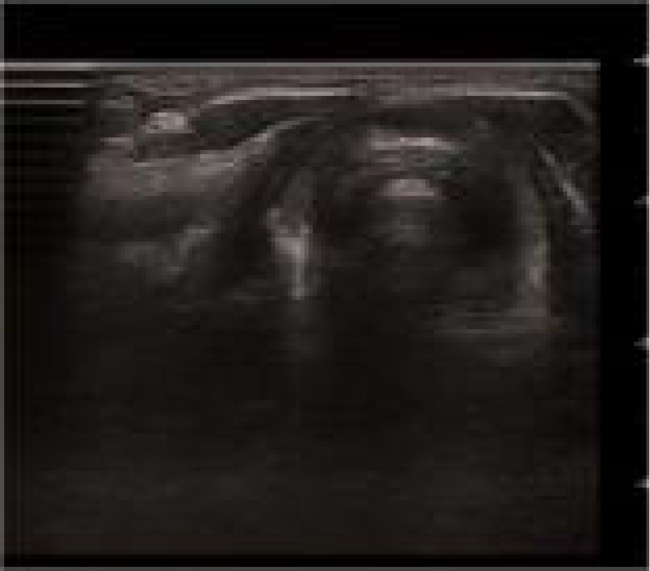
Figure 3
Thyroid ultrasound confirms the absence of thyroid tissue in the normal anatomical location of the neck.
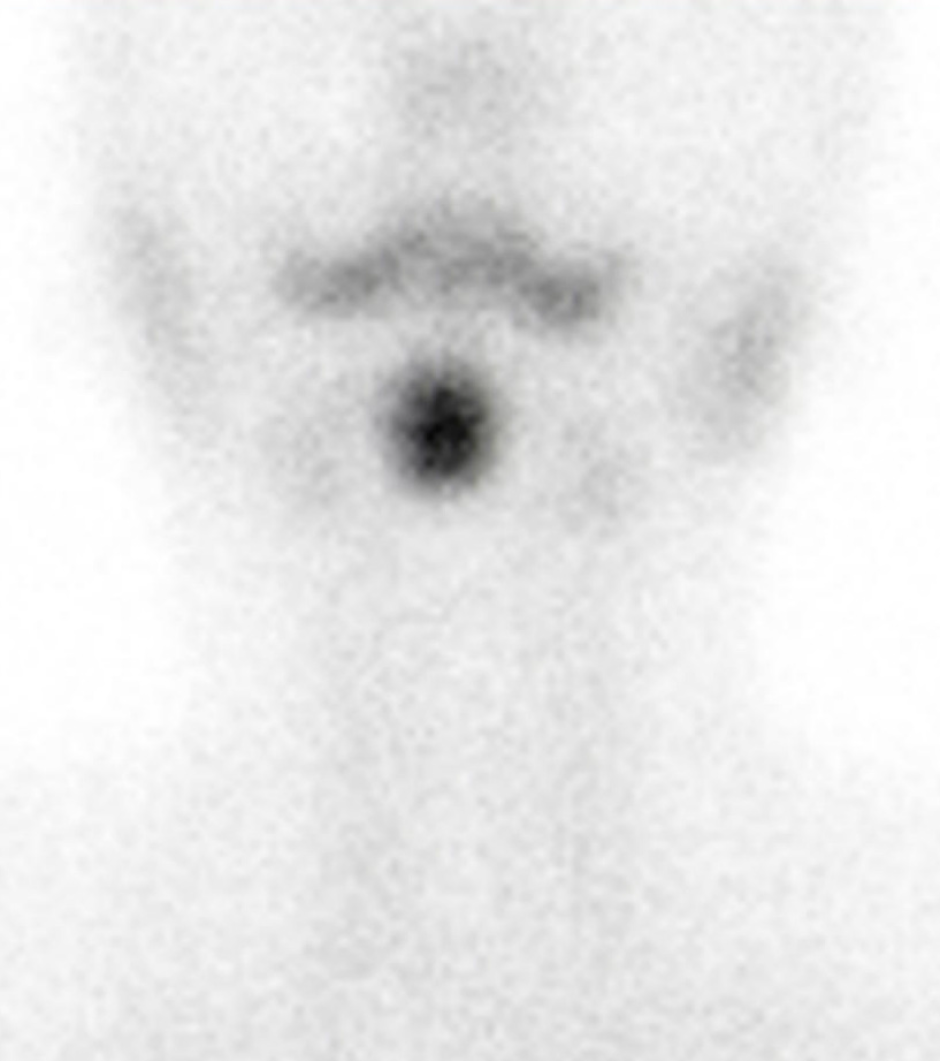
Figure 4
Tc-99 Thyroid scan showing isotopic uptake at the base of the tongue.
Figure 5
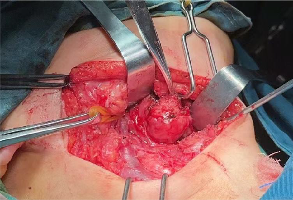
Intraoperative exposure of a tongue root mass, globular in shape.
Figure 6
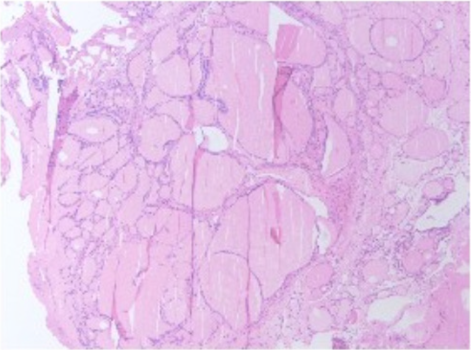
Postoperative pathological findings suggesting well-differentiated thyroid tissue.
- Citation: Yin YT, Gui C. Transposition of the lingual thyroid gland to the submandibular region through a submandibular approach: A case report. World J Clin Cases 2024; 12(21): 4794-4801
- URL: https://www.wjgnet.com/2307-8960/full/v12/i21/4794.htm
- DOI: https://dx.doi.org/10.12998/wjcc.v12.i21.4794


