Copyright
©The Author(s) 2024.
World J Clin Cases. Jul 26, 2024; 12(21): 4783-4788
Published online Jul 26, 2024. doi: 10.12998/wjcc.v12.i21.4783
Published online Jul 26, 2024. doi: 10.12998/wjcc.v12.i21.4783
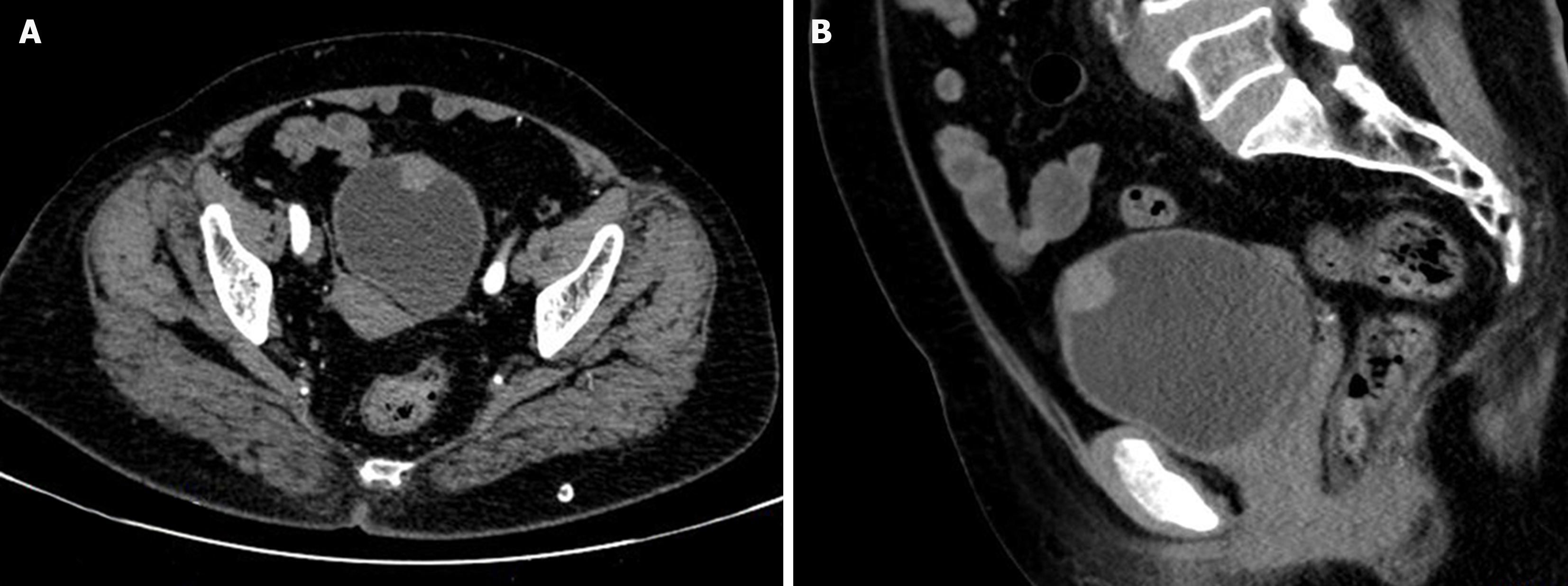
Figure 1 Preoperative pelvic contrast-enhanced computed tomography.
Pelvic contrast-enhanced computed tomography showing a localized tumor on the anterior wall of the bladder measuring approximately 1.68 cm × 1.72 cm × 1.74 cm, which was slightly enhanced. A: Transverse plane; B: Sagittal plane.
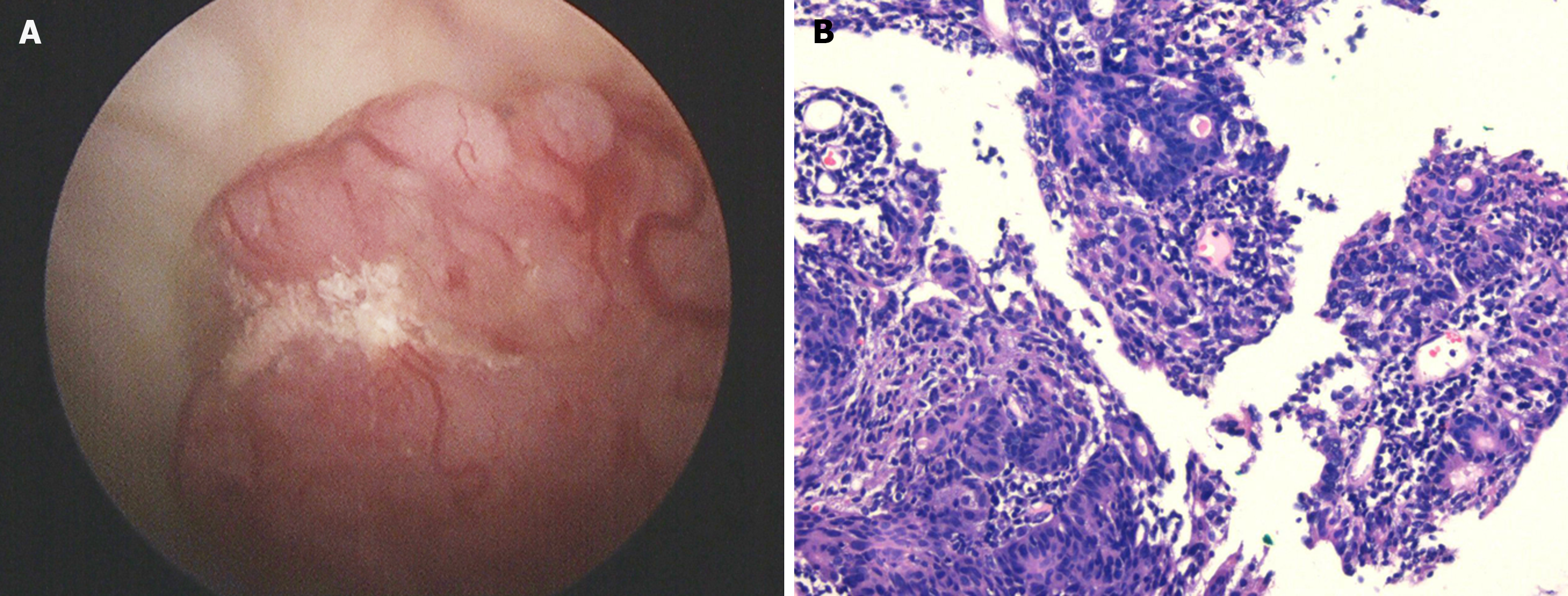
Figure 2 Cystoscopy and pathological examination of tissue samples obtained after biopsy.
A: Cystoscopy showed that there was a 2 cm × 2 cm-sized spherical pink mass on the anterior wall of the bladder with tortuous blood vessels on the surface; B: Pathological examination after biopsy with hematoxylin and eosin staining showed cells with pronounced atypia, large and differently shaped cells and nuclei, and deeply stained nuclei. Bladder malignant tumor was considered after pathological examination (× 200).
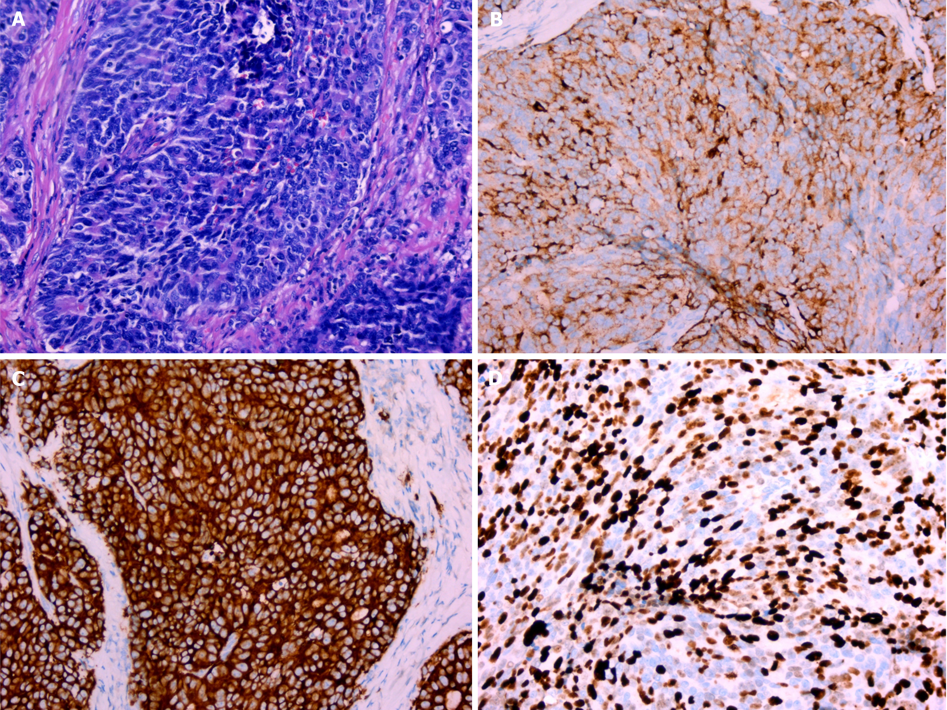
Figure 3 Pathological examination and immunohistochemical staining after surgery.
A: Hematoxylin and eosin staining revealed that the tumor cells had large nuclei, coarse granular chromatin, visible nucleoli, mitotic figures, and palisade-like structures (× 200); B: Chromogranin A was positive (× 200); C: Synaptophysin was positive (× 200); D: The Ki67 index was 70% (× 200).
- Citation: Bai LL, Guo YX, Song SY, Li R, Jiang YQ. Primary large cell neuroendocrine carcinoma of the bladder: A case report. World J Clin Cases 2024; 12(21): 4783-4788
- URL: https://www.wjgnet.com/2307-8960/full/v12/i21/4783.htm
- DOI: https://dx.doi.org/10.12998/wjcc.v12.i21.4783


