Copyright
©The Author(s) 2024.
World J Clin Cases. Jul 26, 2024; 12(21): 4770-4776
Published online Jul 26, 2024. doi: 10.12998/wjcc.v12.i21.4770
Published online Jul 26, 2024. doi: 10.12998/wjcc.v12.i21.4770
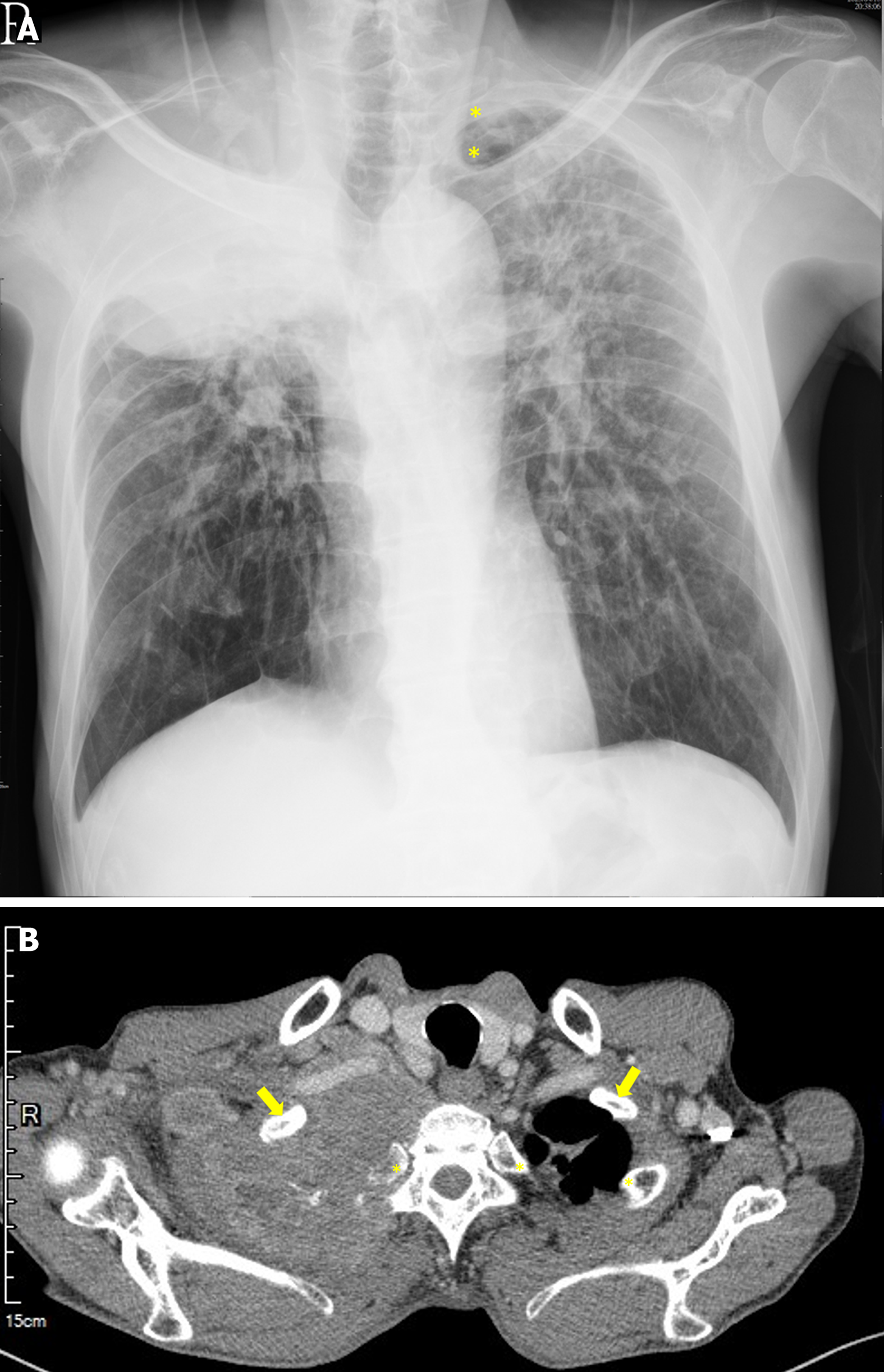
Figure 1 Preoperative imaging shows a huge space-occupying lesion at the right apical lung with adjunct bony destruction.
A: Chest radiograph. The asterisks indicate the second and third ribs at the left side, which was blurred at the right side; B: Chest computed tomography. A soft tissue mass (> 10 cm in diameter) occupied the entire right apical lung. The arrows indicate the first rib. The first rib in the right side was surrounded by the mass. The asterisk indicates the second rib. The second rib in the right side was damaged. The feature of the image highly suggested a locally advanced bronchogenic carcinoma.
Figure 2 Pathological examination of the patient’s lung, which was obtained from the computed tomography-guided biopsy.
A: Lung parenchyma with infiltrating adenocarcinoma cells; B: Infiltrating adenocarcinoma cells with an acinar growth pattern.
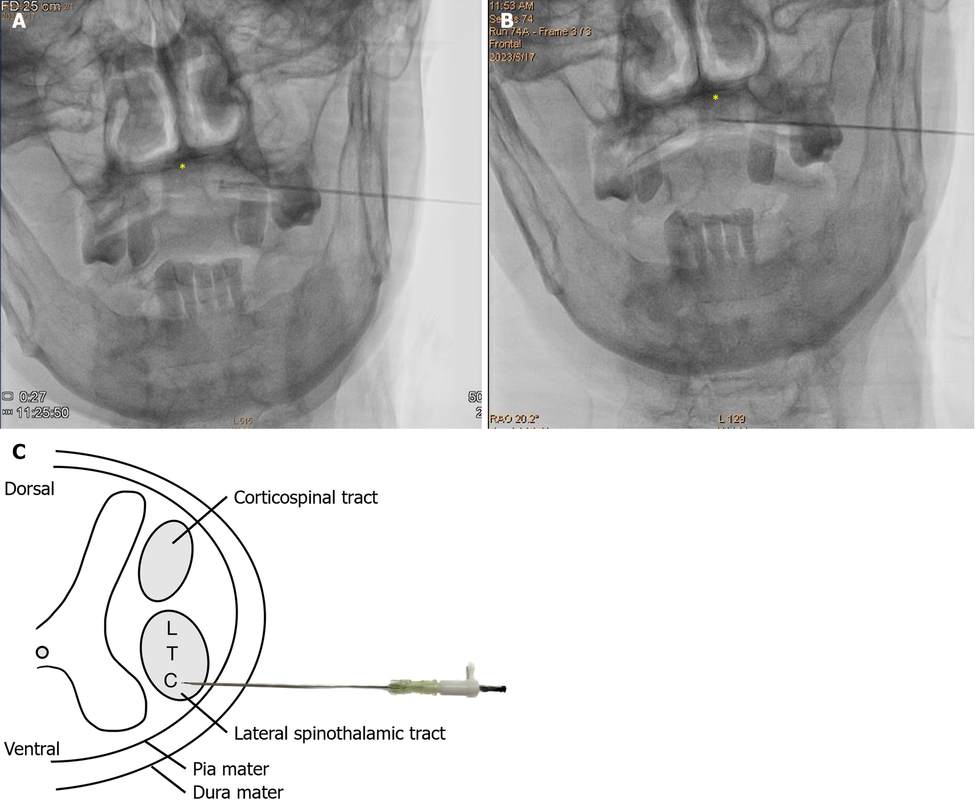
Figure 3 Fluoroscopy image of the anterior-posterior view when advancing the spinal needle during percutaneous cervical cordotomy.
A: When the needle tip enters the intrathecal space, the cerebrospinal fluid is drained. The needle tip did not cross through the midline, which is indicated by an asterisk, the dens of axis (C2); B: Radiofrequency (RF) generator was inserted into the spinal needle. The patient’s discomfort area was triggered by the sensory test. After confirmation of the target lesion, RF thermocoagulation was applied to destroy the neuropathway of the pain; C: Schematic illustration of percutaneous cervical cordotomy and the relative anatomical position of the lateral spinothalamic tract. C: Cervical; L: Lumbar; T: Thoracic.
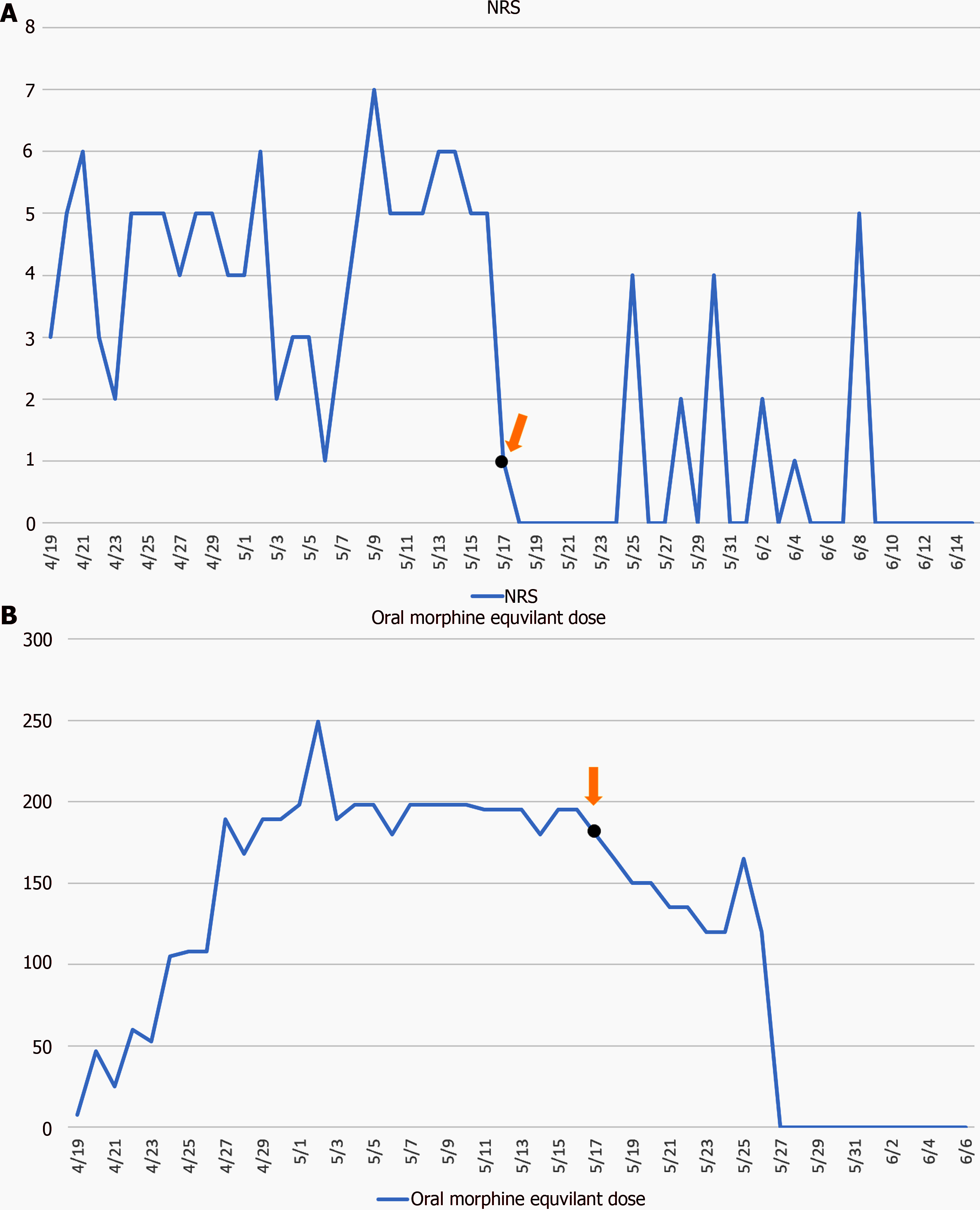
Figure 4 Trajectory of pain alteration.
The orange arrow and black dot represent the percutaneous cervical cordotomy. A: Numeric rating scale score of pain during hospitalization. After the procedure, a pain-free period was noted. He complained of intermittent pain, which can be relieved by nonopioid medications, during the cancer treatment; B: Oral morphine equivalent dosage during hospitalization. After the procedure, the opioid dosage was tapered gradually.
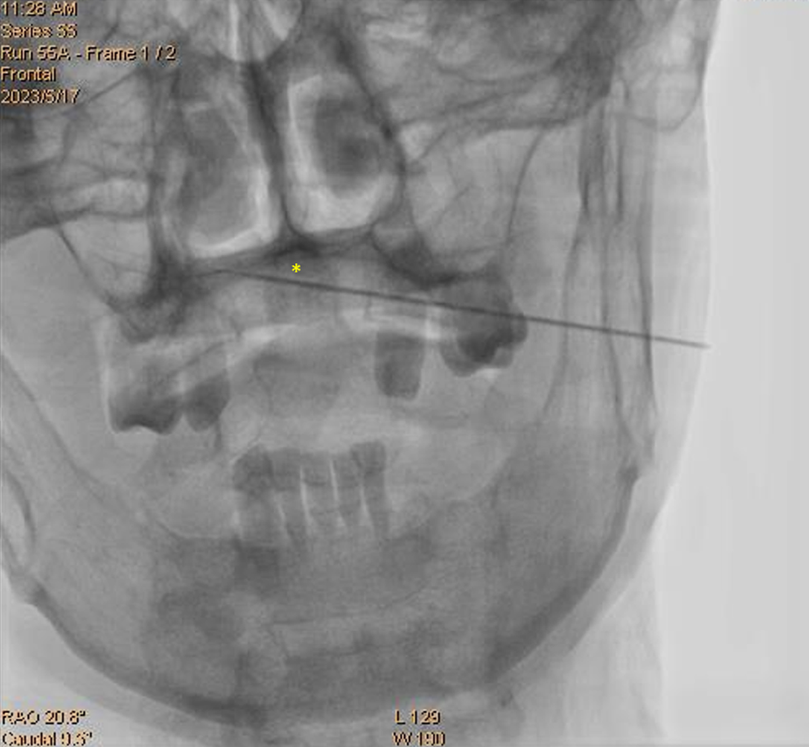
Figure 5 Spinal needle crossing the midline.
The spinal needle appears to have crossed the midline (asterisk) and near the contralateral lamina. However, the patient did not complain of any discomfort. We presumed that the needle may have pushed the spinal cord, instead of being stabbed, when we were advancing the needle. Therefore, the path of the spinal needle should be adjusted after going backward.
- Citation: Lu KY, Lin FS, Lin CS, Lao HC. Percutaneous cervical cordotomy for managing refractory pain in a patient with a Pancoast tumor: A case report. World J Clin Cases 2024; 12(21): 4770-4776
- URL: https://www.wjgnet.com/2307-8960/full/v12/i21/4770.htm
- DOI: https://dx.doi.org/10.12998/wjcc.v12.i21.4770


