Copyright
©The Author(s) 2024.
World J Clin Cases. Jul 16, 2024; 12(20): 4434-4439
Published online Jul 16, 2024. doi: 10.12998/wjcc.v12.i20.4434
Published online Jul 16, 2024. doi: 10.12998/wjcc.v12.i20.4434
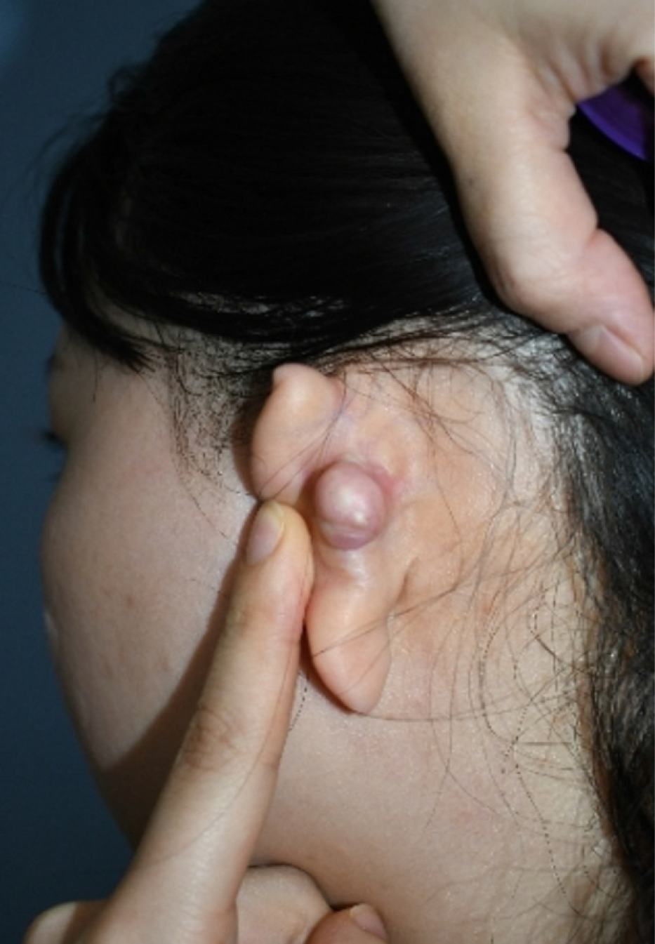
Figure 1 Keloid scar on the posterior side of the left ear.
A 25-year-old woman presented with a keloid scar measuring 2 cm × 2 cm × 1.5 cm on the posterior side of her left ear.
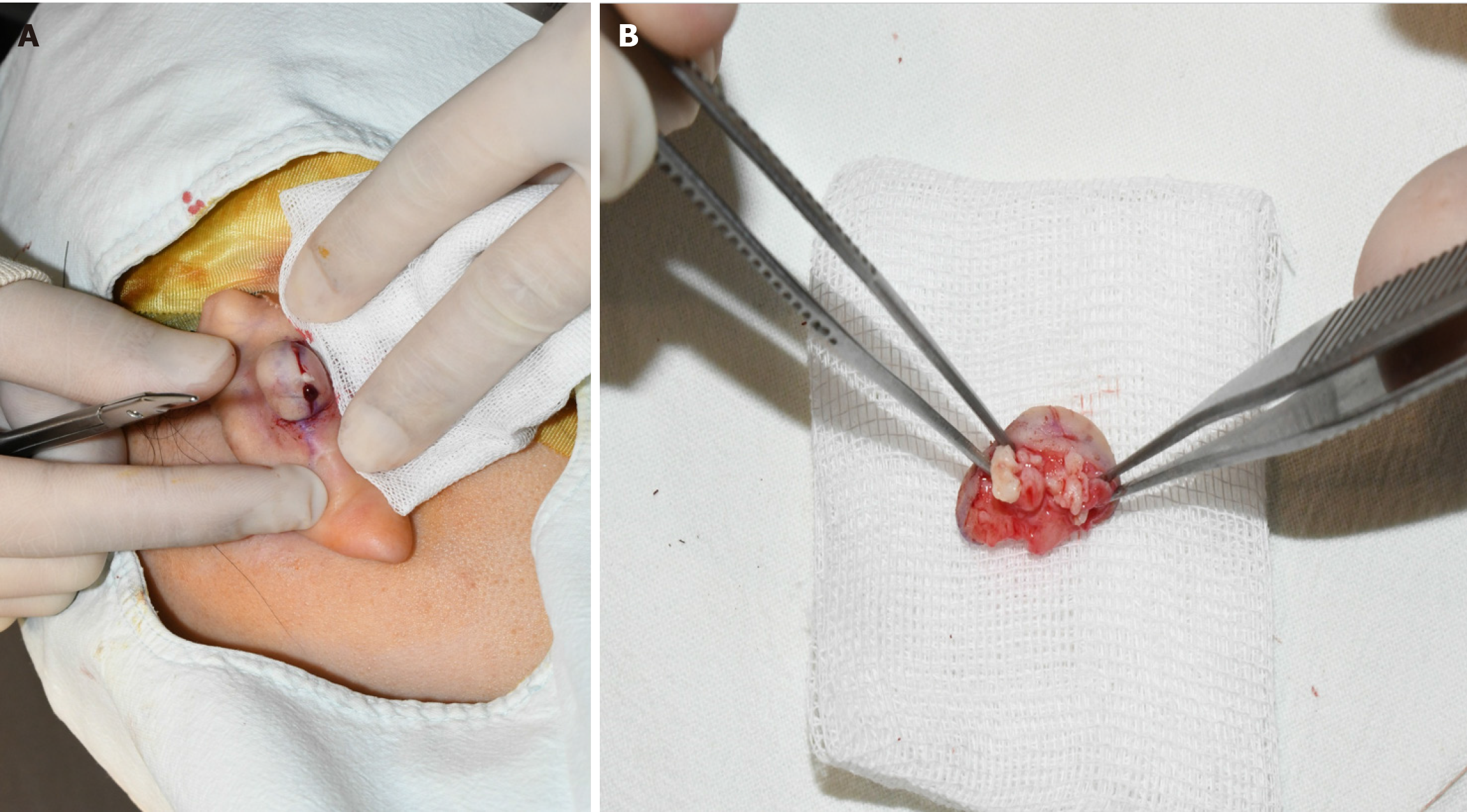
Figure 2 Intraoperative view of the ear keloid containing an epidermal cyst.
A: Rupture of the epidermal cyst upon incision; B: Keloid tissue containing an encapsulated epidermal cyst.
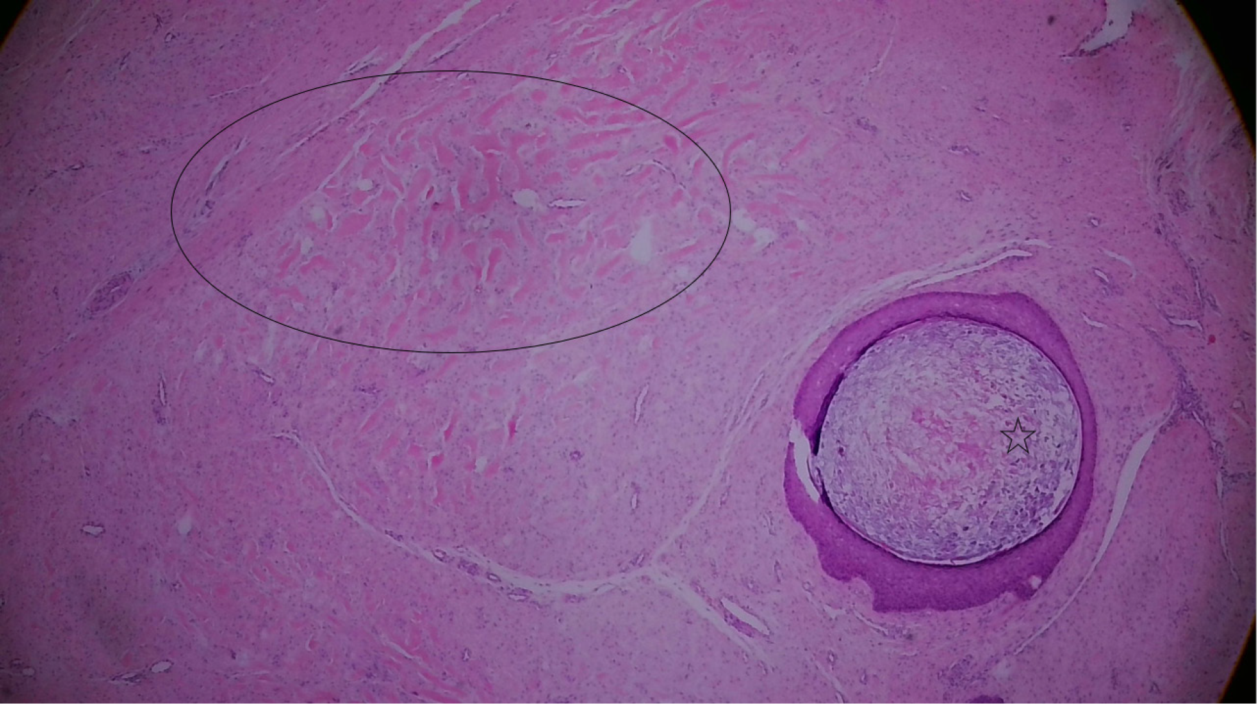
Figure 3 Histological characteristics of the keloid with adjacent epidermal cyst.
Histological image showing thick collagen bundles, a characteristic feature of keloid tissue, marked with circles. The adjacent area is encapsulated by stratified squamous epithelium containing keratin, indicative of an epidermal cyst, marked with asterisks. The tissue is stained with hematoxylin and eosin.
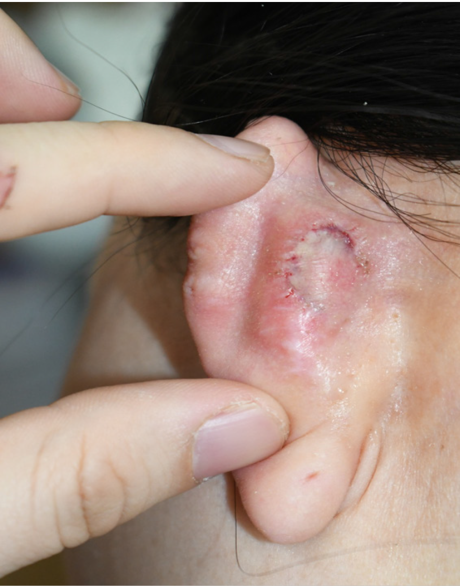
Figure 4
Follow-up photograph taken during suture removal on postoperative day 9.
- Citation: Kim JM, Cheon JS, Choi WY. Ear keloid and epidermal cyst following auricular cartilage harvest for rhinoplasty: A case report. World J Clin Cases 2024; 12(20): 4434-4439
- URL: https://www.wjgnet.com/2307-8960/full/v12/i20/4434.htm
- DOI: https://dx.doi.org/10.12998/wjcc.v12.i20.4434


