Copyright
©The Author(s) 2024.
World J Clin Cases. Jul 16, 2024; 12(20): 4419-4426
Published online Jul 16, 2024. doi: 10.12998/wjcc.v12.i20.4419
Published online Jul 16, 2024. doi: 10.12998/wjcc.v12.i20.4419
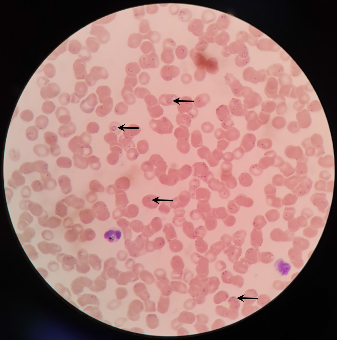
Figure 1 A peripheral blood smear reveals a characteristic early-stage plasmodium falciparum trophozoite.
The blood smear was stained using the Richter Jimsa staining method, and the microscope was set at a magnification of 1000 ×. The arrows represent the plasmodium.
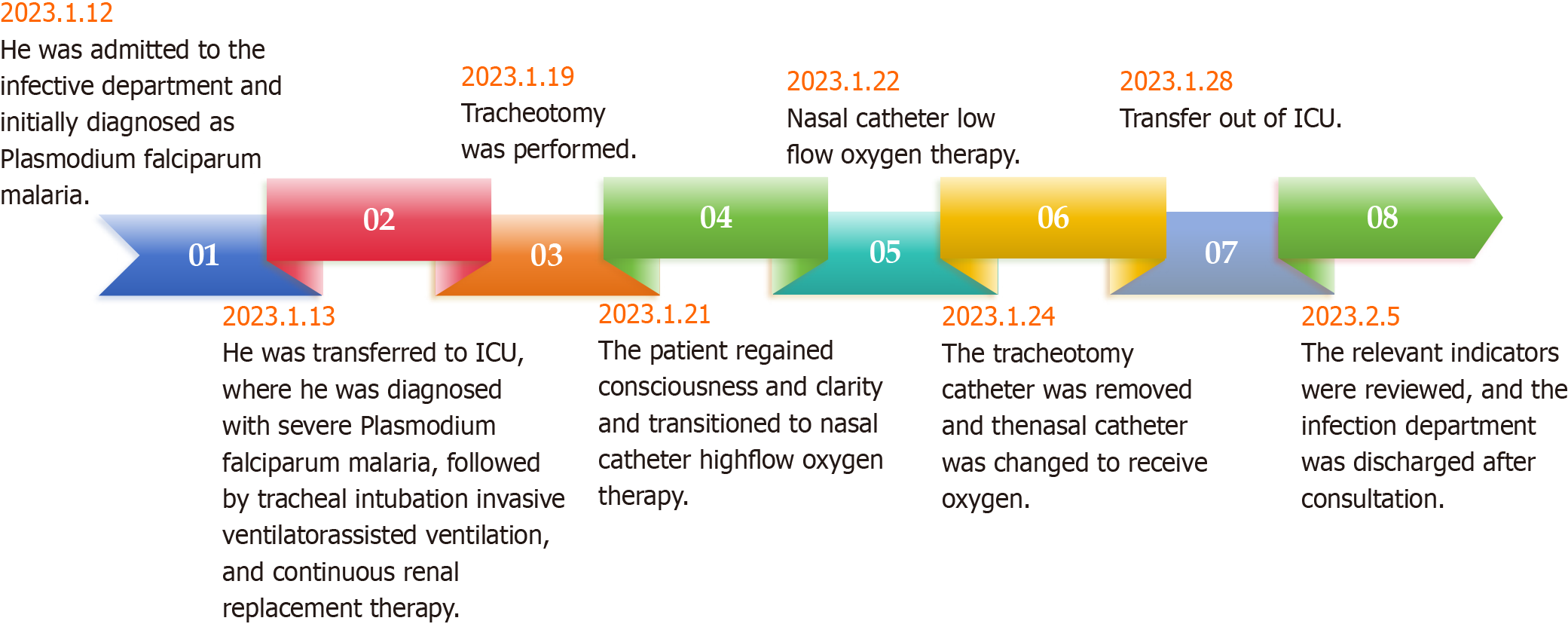
Figure 2 Flow chart of patient treatment.
ICU: Intensive care unit.
- Citation: Zhu YF, Xia WJ, Liu W, Xie JM. Treatment of a patient with severe cerebral malaria during the COVID-19 pandemic in China: A case report. World J Clin Cases 2024; 12(20): 4419-4426
- URL: https://www.wjgnet.com/2307-8960/full/v12/i20/4419.htm
- DOI: https://dx.doi.org/10.12998/wjcc.v12.i20.4419


