Copyright
©The Author(s) 2024.
World J Clin Cases. Jul 16, 2024; 12(20): 4337-4347
Published online Jul 16, 2024. doi: 10.12998/wjcc.v12.i20.4337
Published online Jul 16, 2024. doi: 10.12998/wjcc.v12.i20.4337
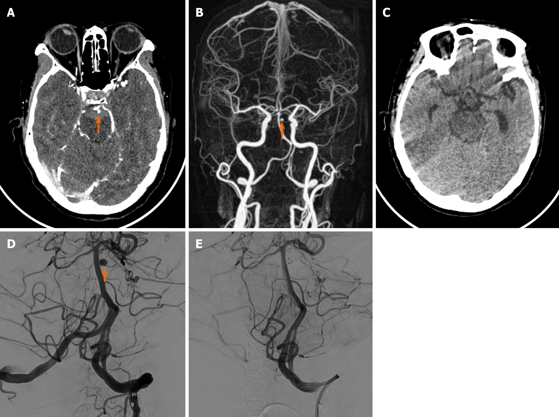
Figure 1 Case 1.
A: Axial enhanced brain computed tomography of a 58-year-old female showed a tiny aneurysm found in the left superior cerebellar artery. Subarachnoid hemorrhage was noted; B: Three-dimensional reconstruction showed a 4.2 mm × 3.0 mm × 2.6 mm aneurysm found in the left superior cerebellar artery; C: Follow-up brain computed tomography (24 months post-operation) showed no new subarachnoid hemorrhage; D: Angiography showed the aneurysm noted on the left side of the basilar artery was clearly visualized by the frontal and lateral views; E: A 3.0 mm × 80.0 mm metallic coil was impacted into the aneurysm sac. Follow-up angiogram confirmed complete embolization and parent artery was intact.
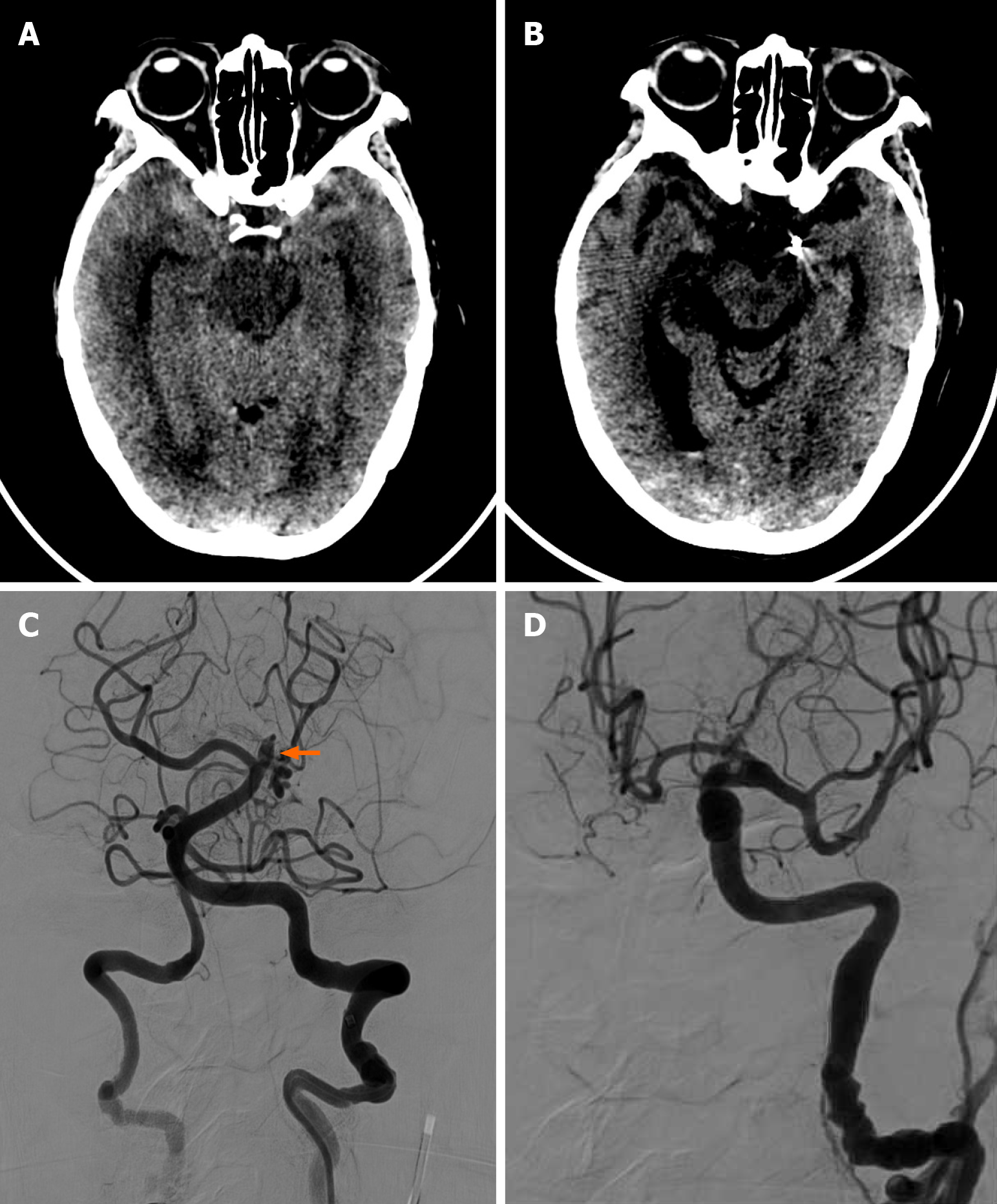
Figure 2 Case 2.
A: Axial brain computed tomography of a 79-year-old female showed bilateral subarachnoid hemorrhage in the bilateral temporal region; B: Axial brain computed tomography post-treatment showed the previous subarachnoid hemorrhage in the bilateral temporal region had subsided. No intracranial hemorrhage was found; C: Angiography showed a bulging aneurysm in the basilar perforated branches; D: Final angiography showed complete embolization of the aneurysm.
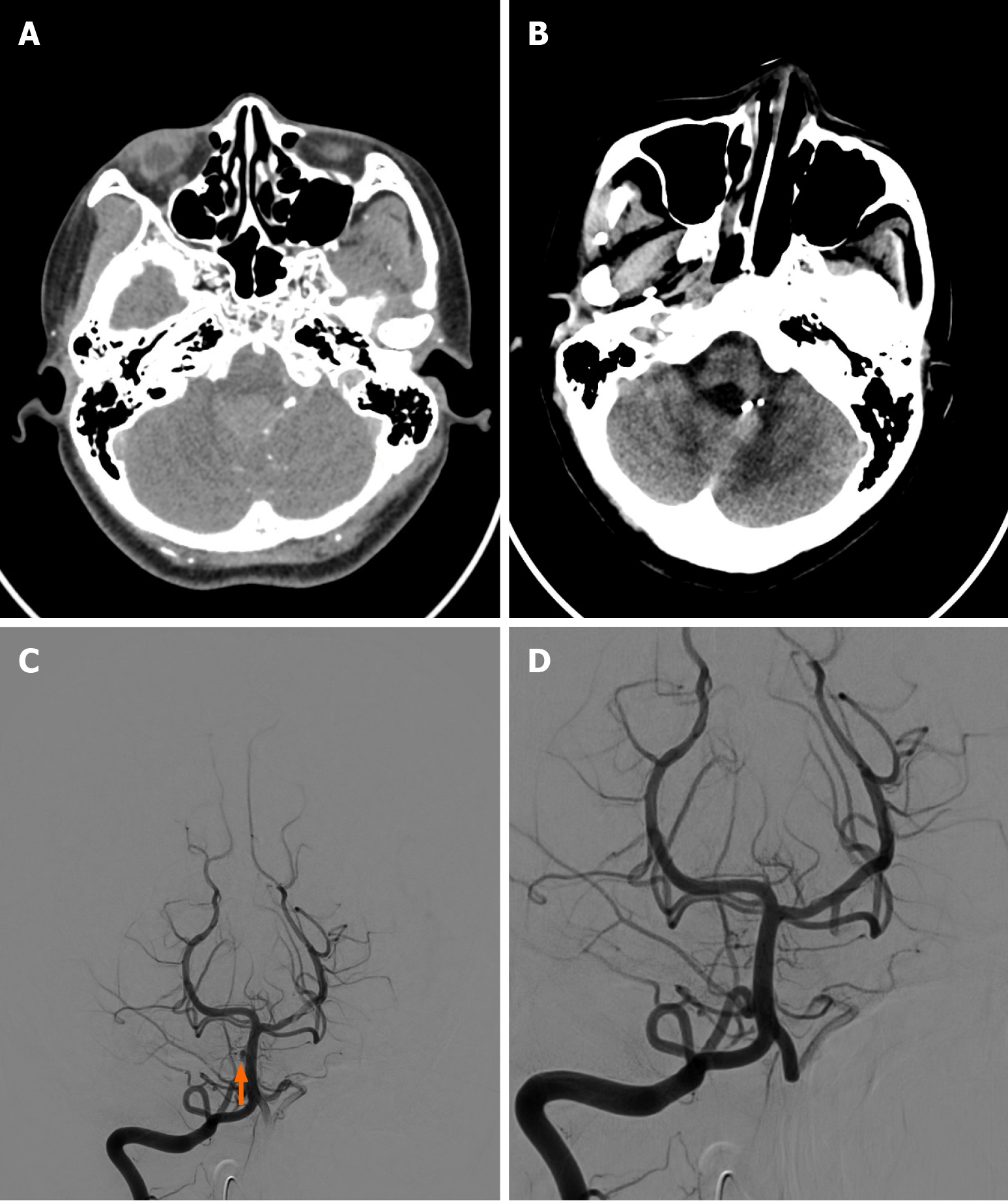
Figure 3 Case 3.
A: Axial brain computed tomography with contrast of a 31-year-old female showed an irregular saccular structure found in one of the left distal vertebral artery meningeal branches; B: Follow-up (24 months) axial brain non-contrast computed tomography of the same patient showed the previous subarachnoid hemorrhage regressed, and no new subarachnoid hemorrhage was noted; C: Angiography showed a tiny semi-circle saccular lesion noted on the right side of the basilar artery, and it was considered an unruptured basilar artery perforator aneurysm; D: Metallic coil embolization of these aneurysms was performed.
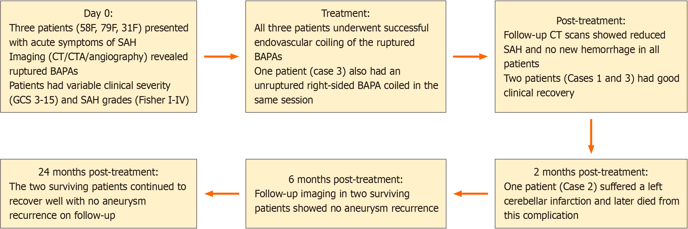
Figure 4 Treatment and follow-up timeline of patients with basilar artery perforator aneurysm.
BAPA: Basilar artery perforator aneurysm; CT: Computed tomography; CTA: Computed tomography angiography; F: Female; GCS: Glasgow Coma Score; SAH: Subarachnoid hemorrhage.
- Citation: Man IC, Pan TM, U KC. An unusual etiology of subarachnoid hemorrhage, basilar artery perforator aneurysms, in Macao: Three case reports and review of literature. World J Clin Cases 2024; 12(20): 4337-4347
- URL: https://www.wjgnet.com/2307-8960/full/v12/i20/4337.htm
- DOI: https://dx.doi.org/10.12998/wjcc.v12.i20.4337


