Copyright
©The Author(s) 2024.
World J Clin Cases. Jul 16, 2024; 12(20): 4317-4324
Published online Jul 16, 2024. doi: 10.12998/wjcc.v12.i20.4317
Published online Jul 16, 2024. doi: 10.12998/wjcc.v12.i20.4317
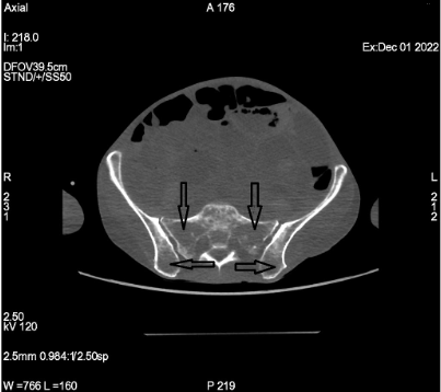
Figure 1 Osteolytic lesions in pelvic bones (arrows) in computed tomography scan.
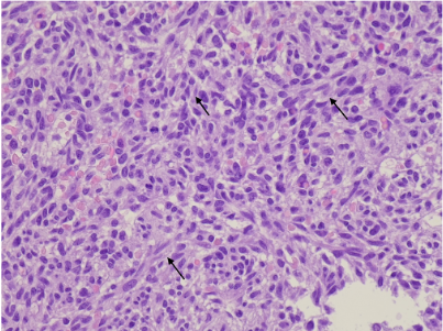
Figure 2 Hematoxylin-eosin staining of trephine biopsy presenting with mast cell leukemia infiltrates.
Atypical mast cell morphology–spindle-shaped cells (magnification × 400).
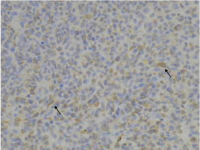
Figure 3 Mast cell tryptase immunostaining in bone marrow infiltrates (magnification × 400).
Figure 4 Aberrant diffuse CD25 expression in neoplastic mast cells in trephine biopsy (magnification × 200).
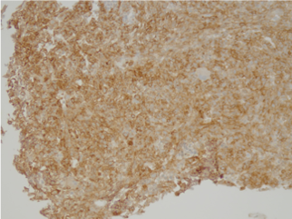
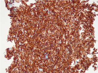
Figure 5 Membrane CD117 expression in neoplastic mast cells in trephine biopsy (magnification × 200).
- Citation: Wysocki MT, Gonciarz M, Puła B. Treatment refractory mast cell leukemia with dominant gastrointestinal manifestation and concomitant skin symptoms: A case report. World J Clin Cases 2024; 12(20): 4317-4324
- URL: https://www.wjgnet.com/2307-8960/full/v12/i20/4317.htm
- DOI: https://dx.doi.org/10.12998/wjcc.v12.i20.4317


