Copyright
©The Author(s) 2024.
World J Clin Cases. Jan 16, 2024; 12(2): 451-459
Published online Jan 16, 2024. doi: 10.12998/wjcc.v12.i2.451
Published online Jan 16, 2024. doi: 10.12998/wjcc.v12.i2.451
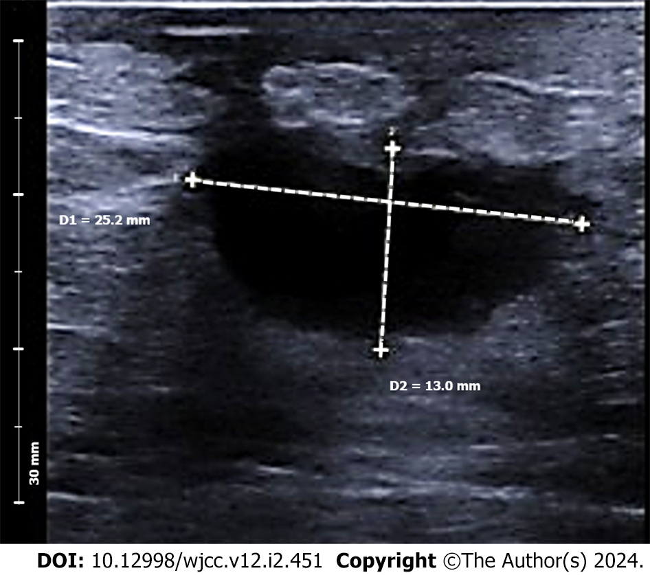
Figure 1 Thickening of subcutaneous tissue, located at 11-12 o'clock position in the right breast (suspected to be an inflammatory lesion with localized inflammatory edema of the subcutaneous tissue), measuring up to 8.
7 mm in thickness. Adjacent to the nipple is a hypoechoic nodule, measuring 25 mm × 13 mm, situated 22 mm deep to the body surface, with well-defined borders, regular shape, normal aspect ratio, homogenous internal echogenicity, no nodule calcification, unchanged posterior echogenicity, and peripheral dotted lines indicative of blood flow signal. The gland thickness surrounding the mass is approximately 2 cm.
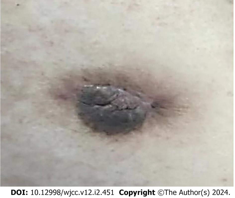
Figure 2 Sizable mass in the retro-areolar area of the right breast, characterized by skin erythema and nipple inversion.
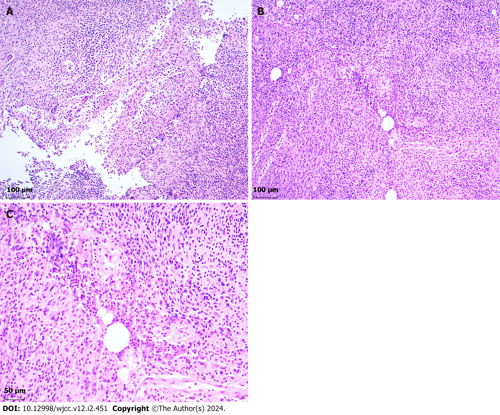
Figure 3 Pathological investigation revealed a poorly formed non-caseating granuloma.
A: Depicting the poorly formed non-caseating granuloma encircled by epithelioid cells, lymphocytes, plasma cells, neutrophils, and multinucleate giant cells. [hematoxylin and eosin (HE), 100 × magnification]; B: Illustrating the non-caseating granuloma centered on a vacuolated space (HE, 100 × magnification); C: High-resolution view of Figure 3B, highlighting the vacuolated space rimmed by neutrophils and surrounded by epithelioid cells, lymphocytes, plasma cells, and multinucleate giant cells (HE, 200 × magnification).
Figure 4 Depicting the well-healed surgical incision three months postoperatively.
- Citation: Cui LY, Sun CP, Li YY, Liu S. Granulomatous mastitis in a 50-year-old male: A case report and review of literature. World J Clin Cases 2024; 12(2): 451-459
- URL: https://www.wjgnet.com/2307-8960/full/v12/i2/451.htm
- DOI: https://dx.doi.org/10.12998/wjcc.v12.i2.451


