Copyright
©The Author(s) 2024.
World J Clin Cases. Jan 16, 2024; 12(2): 425-430
Published online Jan 16, 2024. doi: 10.12998/wjcc.v12.i2.425
Published online Jan 16, 2024. doi: 10.12998/wjcc.v12.i2.425
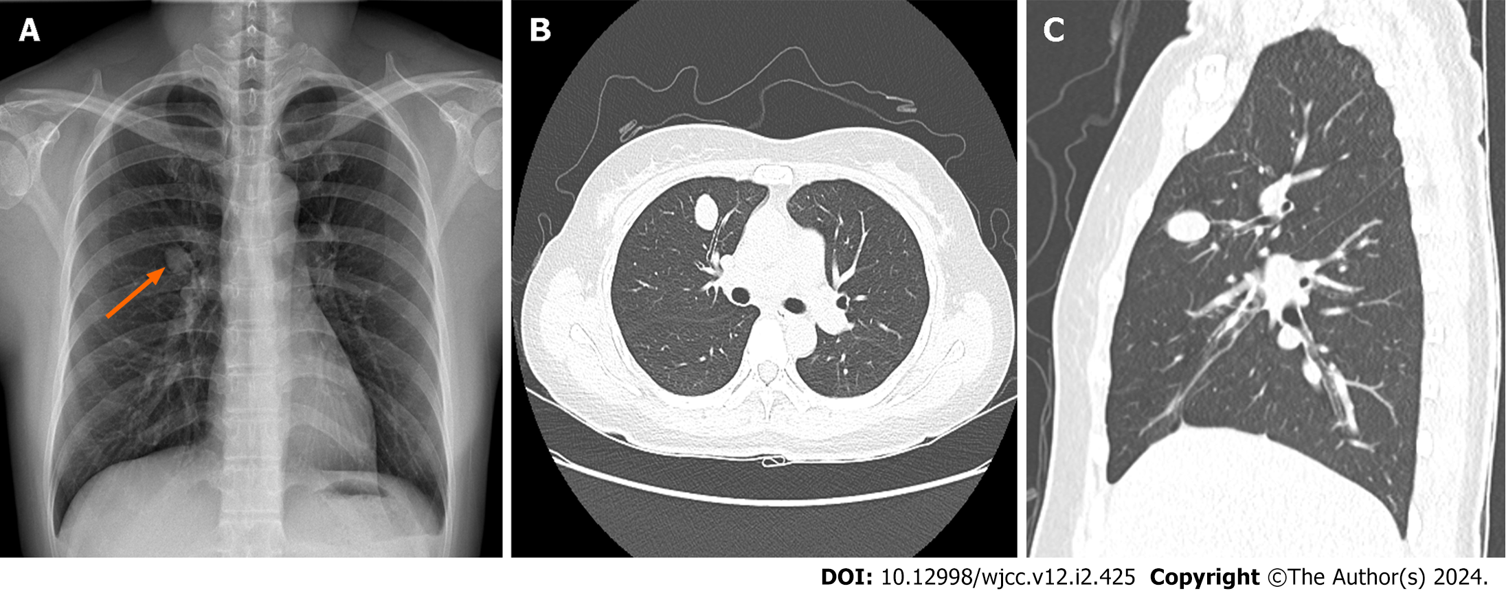
Figure 1 Preoperative imaging studies.
A: Chest radiograph showing a solitary pulmonary nodule of the right hilar area (orange arrow); B and C: Similar findings are observed on chest computed tomography (axial and sagittal views), with the nodule situated in the anterior segment of the right upper lobe.
Figure 2 Operative views of the segmentectomy.
A: A solid, round, and mobile pulmonary nodule is observed; however, there is no minor fissure; B: The anterior segmental artery (A3) and bronchus (B3) are identified (white arrows); C: The resected anterior segment of the right upper lobe, with visible vascular and bronchial stumps (orange arrow).
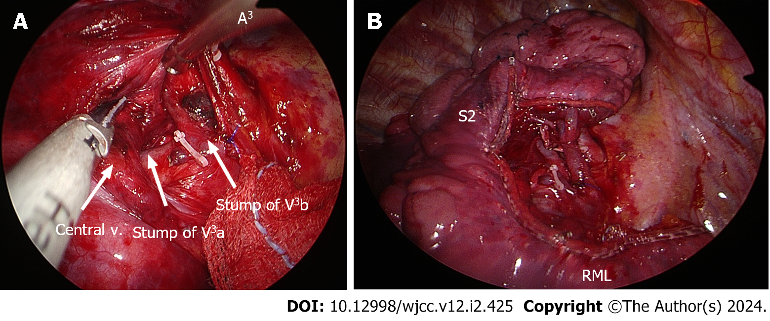
Figure 3 Right upper lobe stumps at the resection line.
A: Anterior segmental veins (V3b and V3a) are clearly visible; B: The surgical edge at the completion of uniportal video-assisted thoracoscopic right upper lobe anterior segmentectomy.
Figure 4 Postoperative findings.
A: A clear upright film (anteroposterior) acquired in the recovery room; B: Image of the gross specimen revealing a well-demarcated, unencapsulated, yellowish-gray mass without necrosis, measuring 2.2 cm × 2.0 cm. This solitary pulmonary nodule was situated in close proximity to the anterior segmental bronchus of the right upper lobe.
Figure 5 Microscopic morphology.
A: Microscopic examination showing a hypercellular nodule comprising spindle-shaped cells arranged in a fascicular growth pattern; B: At higher magnification, plump spindle cell proliferation is observed, interspersed with chronic inflammatory cells; C: Immunohistochemically, the spindle cells exhibit diffuse and strong positivity for anaplastic lymphoma kinase ventana anti-anaplastic lymphoma kinase.
- Citation: Ahn S, Moon Y. Uniportal video-assisted thoracoscopic fissureless right upper lobe anterior segmentectomy for inflammatory myofibroblastic tumor: A case report. World J Clin Cases 2024; 12(2): 425-430
- URL: https://www.wjgnet.com/2307-8960/full/v12/i2/425.htm
- DOI: https://dx.doi.org/10.12998/wjcc.v12.i2.425


