Copyright
©The Author(s) 2024.
World J Clin Cases. Jan 16, 2024; 12(2): 405-411
Published online Jan 16, 2024. doi: 10.12998/wjcc.v12.i2.405
Published online Jan 16, 2024. doi: 10.12998/wjcc.v12.i2.405
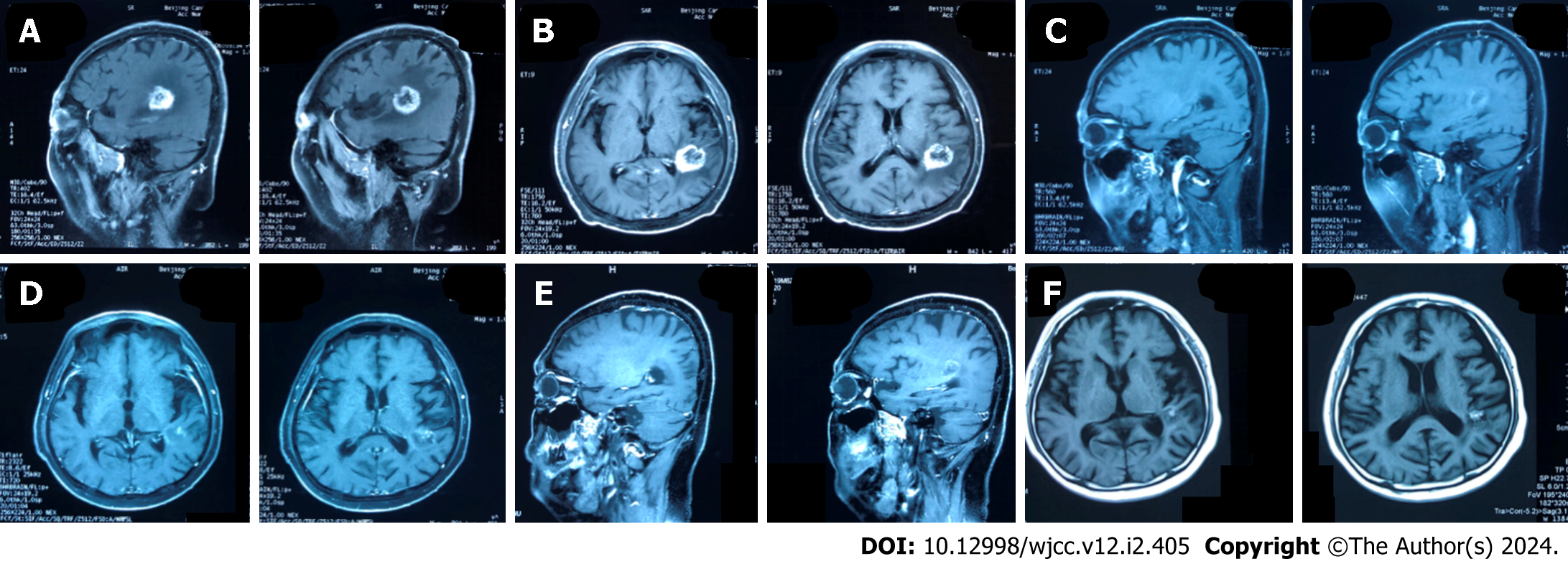
Figure 1 On February 22, 2023, the patient's magnetic resonance imaging showed large metastatic lesions in the left temporal lobe and the upper lobe of the right lung.
In June 2023, the patient came to our hospital to receive two cycles of irinotecan and bevacizumab, and the magnetic resonance imaging (MRI) of the head examined in other hospitals showed a significant shrinkage of intracranial metastases compared to the previous one. In August 2023, the intracranial metastases were again slightly reduced. A and B: Brain MRI examination T1 weighted image in sagittal plane and cross section showing two lumps in the brain parenchyma, February22, 2023; C and D: Head MRI examination T1 weighted image showing intracranial metastases significantly response was seen clinically after 6 chemotherapy in the corresponding position, June, 2023; E and F: The intracranial metastasis was again slightly reduced, late August, 2023.
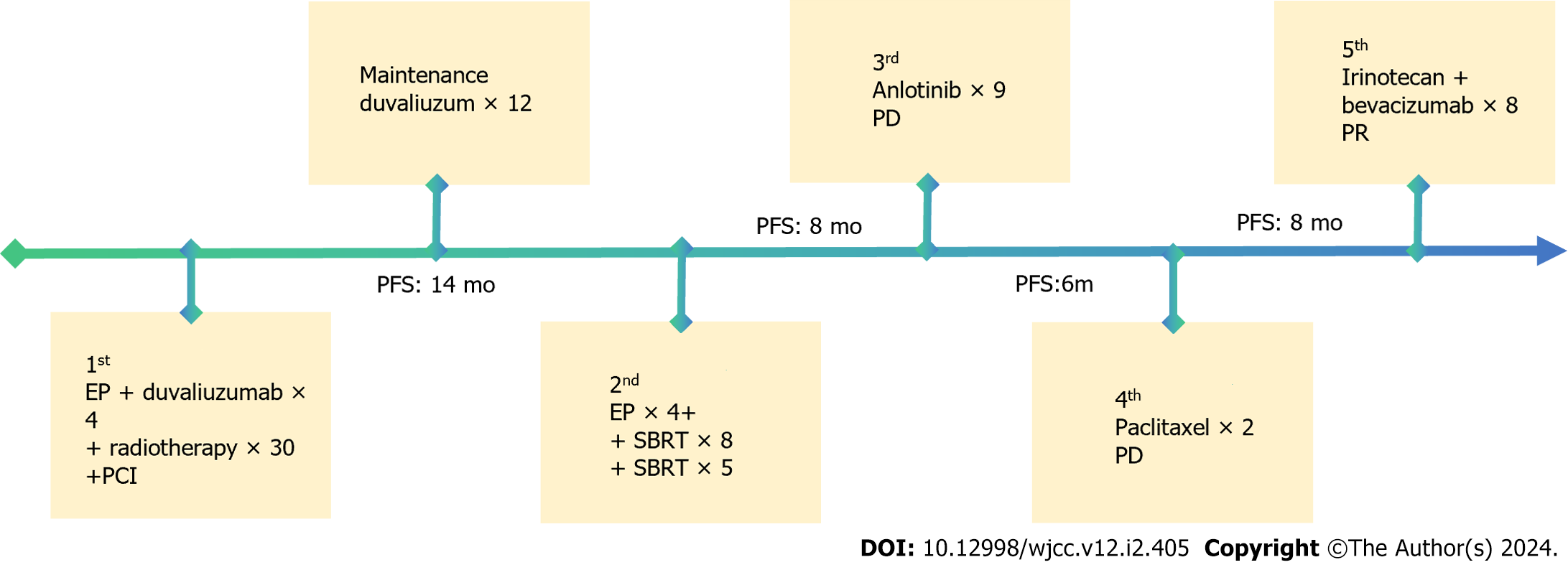
Figure 2 Patient's treatment history.
PD: Progressive disease; PR: Partial response; SBRT: Stereotactic body radiotherapy; PCI: Preventive cranial irradiation; EP: Etoposide plus cisplatin; PFS: Progression-free survival.
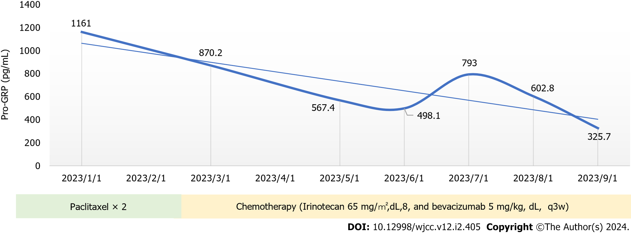
Figure 3 Changes in pro-gastrin-releasing peptide.
Pro-GRP: Pro-gastrin-releasing peptide.
- Citation: Yang HY, Xia YQ, Hou YJ, Xue P, Zhu SJ, Lu DR. Chemotherapy combined with bevacizumab for small cell lung cancer with brain metastases: A case report. World J Clin Cases 2024; 12(2): 405-411
- URL: https://www.wjgnet.com/2307-8960/full/v12/i2/405.htm
- DOI: https://dx.doi.org/10.12998/wjcc.v12.i2.405


