Copyright
©The Author(s) 2024.
World J Clin Cases. Jan 16, 2024; 12(2): 392-398
Published online Jan 16, 2024. doi: 10.12998/wjcc.v12.i2.392
Published online Jan 16, 2024. doi: 10.12998/wjcc.v12.i2.392
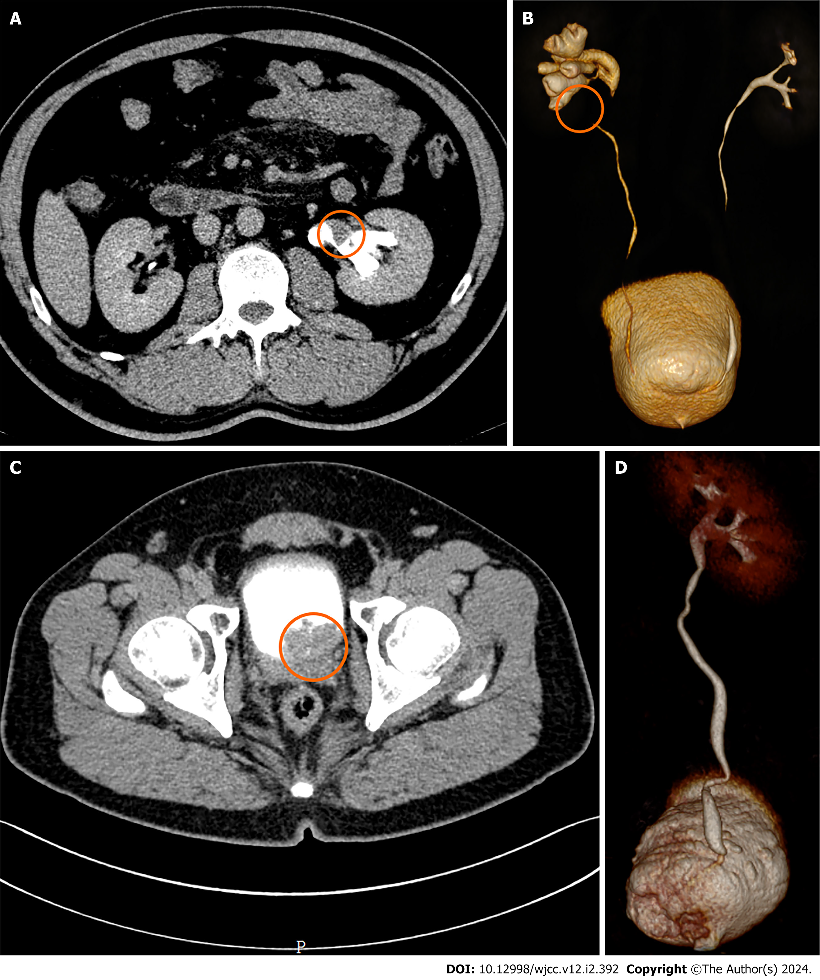
Figure 1 Computed tomography urography of the abdomen.
A: Filling defect from upper segment of left ureter (orange circle); B: A tumor involving the left renal pelvis (orange circle); C: Filling defect from the bladder (orange circle); D: The right urinary tract is unobstructed.
Figure 2 Histopathological findings of cystoscope biopsy and postoperative pathology.
A: Bladder urothelial carcinoma (× 400). B: Tumors were recognized as papillary urothelial carcinoma on the excised bladder (× 400).
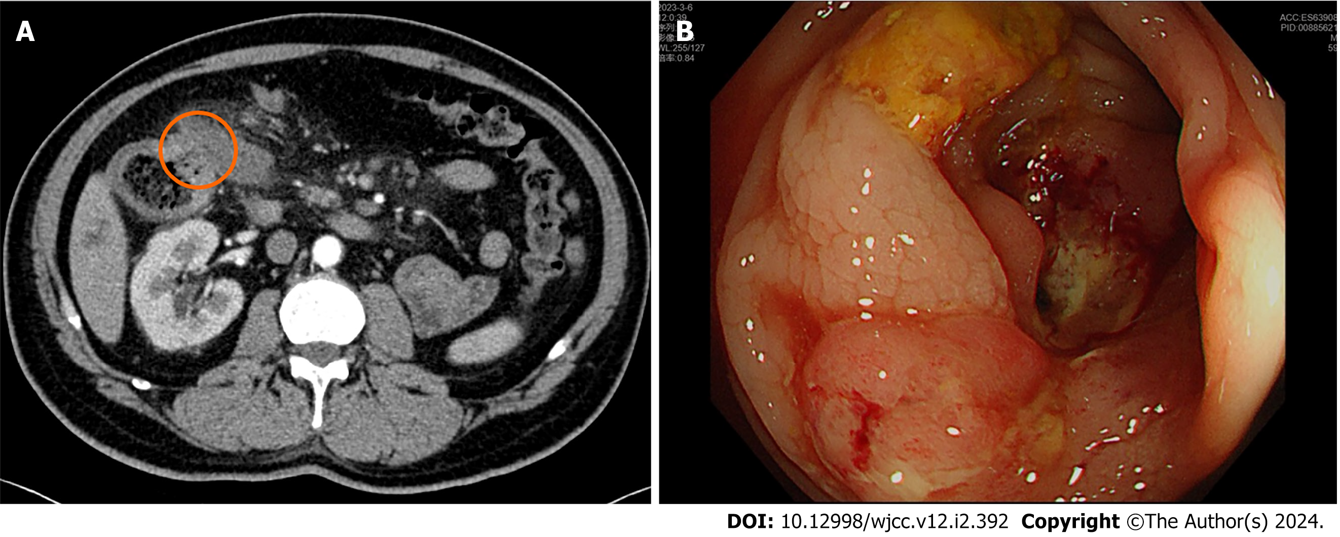
Figure 3 Computed tomography of the abdomen and colonoscopy examination.
A: A tumor of the ascending colon (orange circle); B: A tumor in the descending colon.
Figure 4 Histopathological findings of colonoscopy biopsy and postoperative pathology.
A: Adenocarcinoma of the descending colon (× 400); B: Tumors were recognized as adenocarcinoma on the excised colon (× 400).
- Citation: Chen J, Huang HY, Zhou HC, Liu LX, Kong CF, Zhou Q, Fei JM, Zhu YM, Liu H, Tang YC, Zhou CZ. Three cancers in the renal pelvis, bladder, and colon: A case report. World J Clin Cases 2024; 12(2): 392-398
- URL: https://www.wjgnet.com/2307-8960/full/v12/i2/392.htm
- DOI: https://dx.doi.org/10.12998/wjcc.v12.i2.392


