Copyright
©The Author(s) 2024.
World J Clin Cases. Jan 16, 2024; 12(2): 383-391
Published online Jan 16, 2024. doi: 10.12998/wjcc.v12.i2.383
Published online Jan 16, 2024. doi: 10.12998/wjcc.v12.i2.383
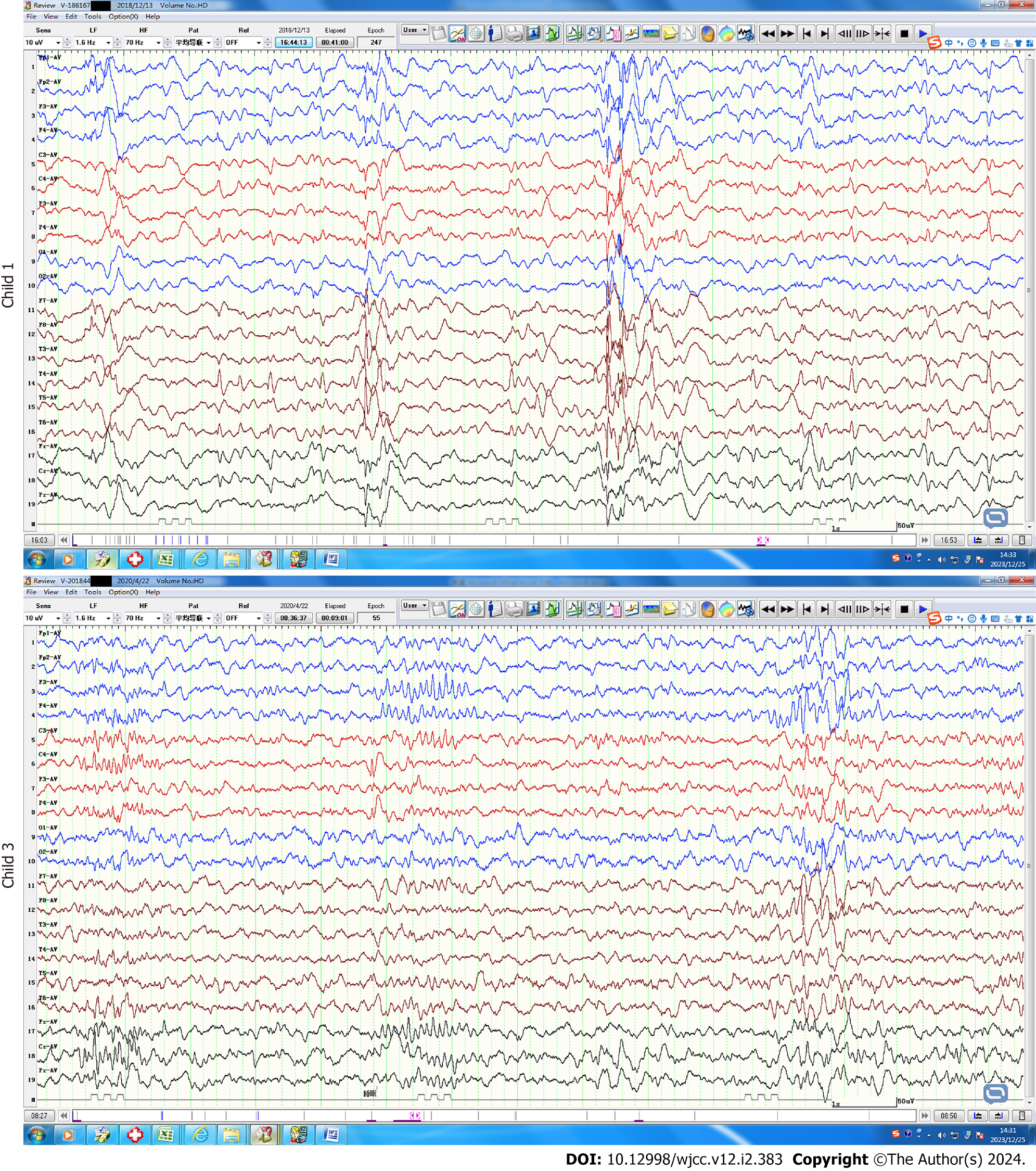
Figure 1 Video electroencephalogram findings.
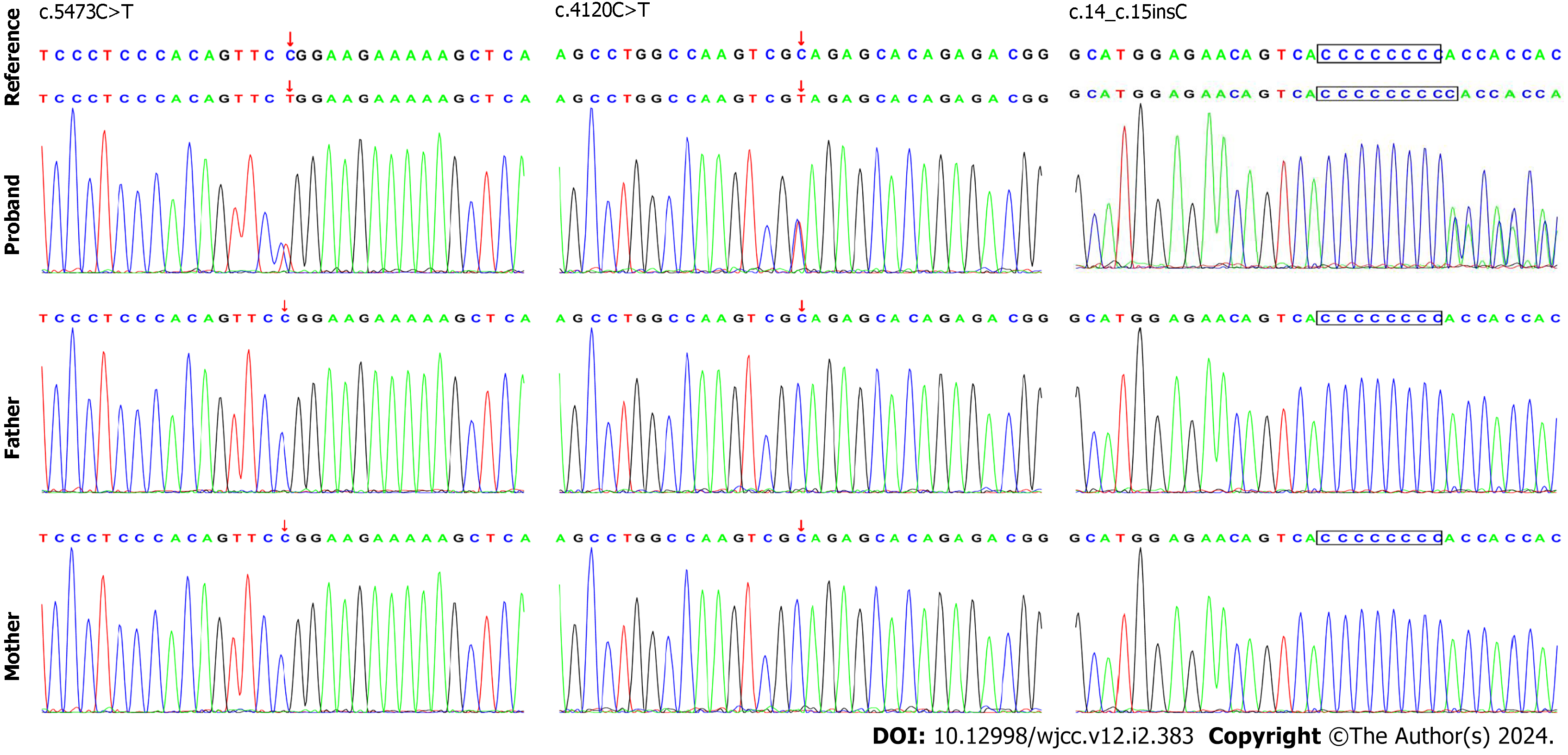
Figure 2 SETD1B gene mutations (red arrow).
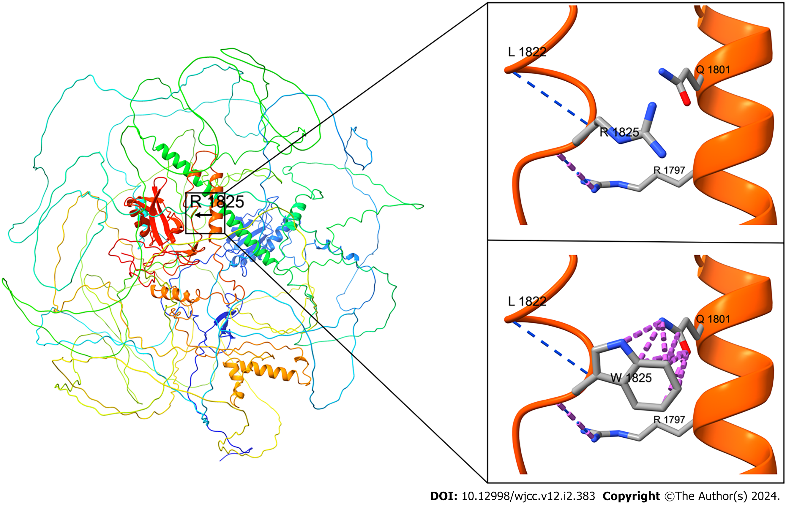
Figure 3 Three-dimensional structure diagram of SETD1B albumen.
Wild-type panorama; the wild-type fragment (Arg1825) has two hydrogen bonds (blue) with Leu1822 and Arg1797, respectively. However, there is an interaction force (repulsion, purple) between the R group atom in the Trp side chain and the R group atom in the Gln1801 side chain after mutation, which is not conducive to interaction and correct protein folding, and may lead to changes in the protein conformation, thus affecting protein function, (left) upper right; mutant local map (bottom right). However, there is no hydrogen bond change before and after mutation.
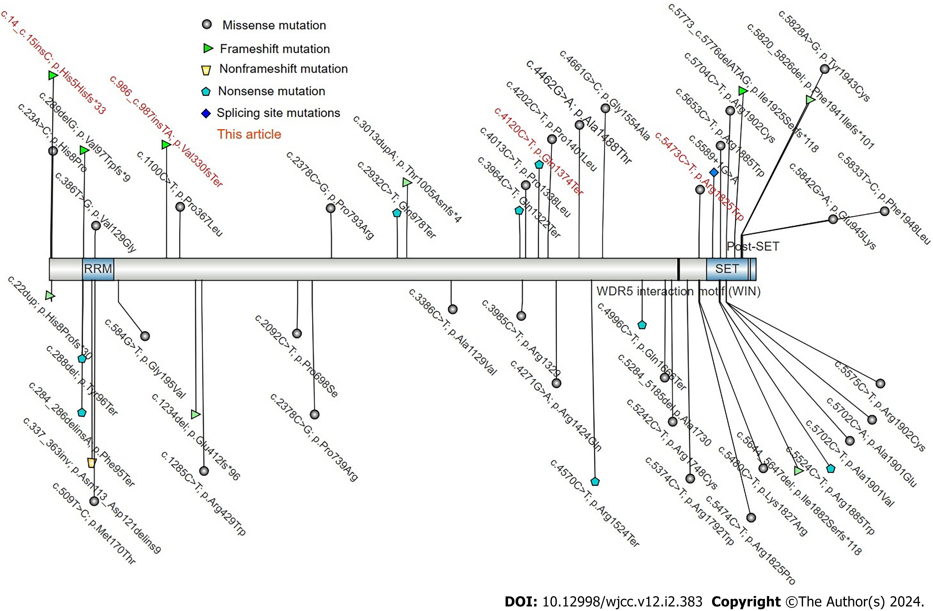
Figure 4 Mutation map of the SETD1B gene (large fragment deletion not shown).
- Citation: Ding L, Wei LW, Li TS, Chen J. Mental retardation, seizures and language delay caused by new SETD1B mutations: Three case reports. World J Clin Cases 2024; 12(2): 383-391
- URL: https://www.wjgnet.com/2307-8960/full/v12/i2/383.htm
- DOI: https://dx.doi.org/10.12998/wjcc.v12.i2.383


