Copyright
©The Author(s) 2024.
World J Clin Cases. Jan 16, 2024; 12(2): 361-366
Published online Jan 16, 2024. doi: 10.12998/wjcc.v12.i2.361
Published online Jan 16, 2024. doi: 10.12998/wjcc.v12.i2.361
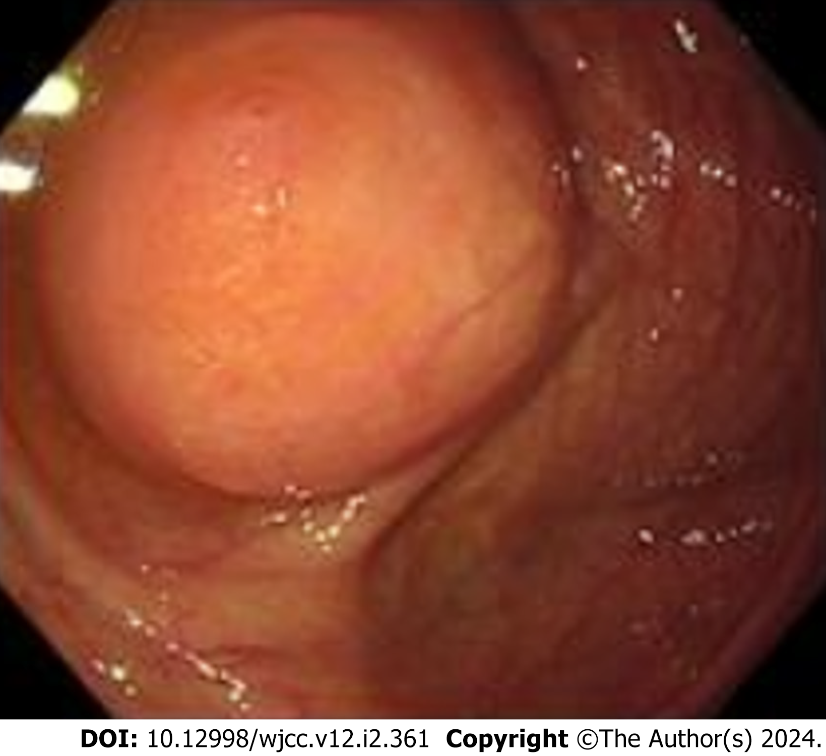
Figure 1 Enteroscopy.
A raised mass with a smooth surface measuring 3.0 cm was observed in the cecum.
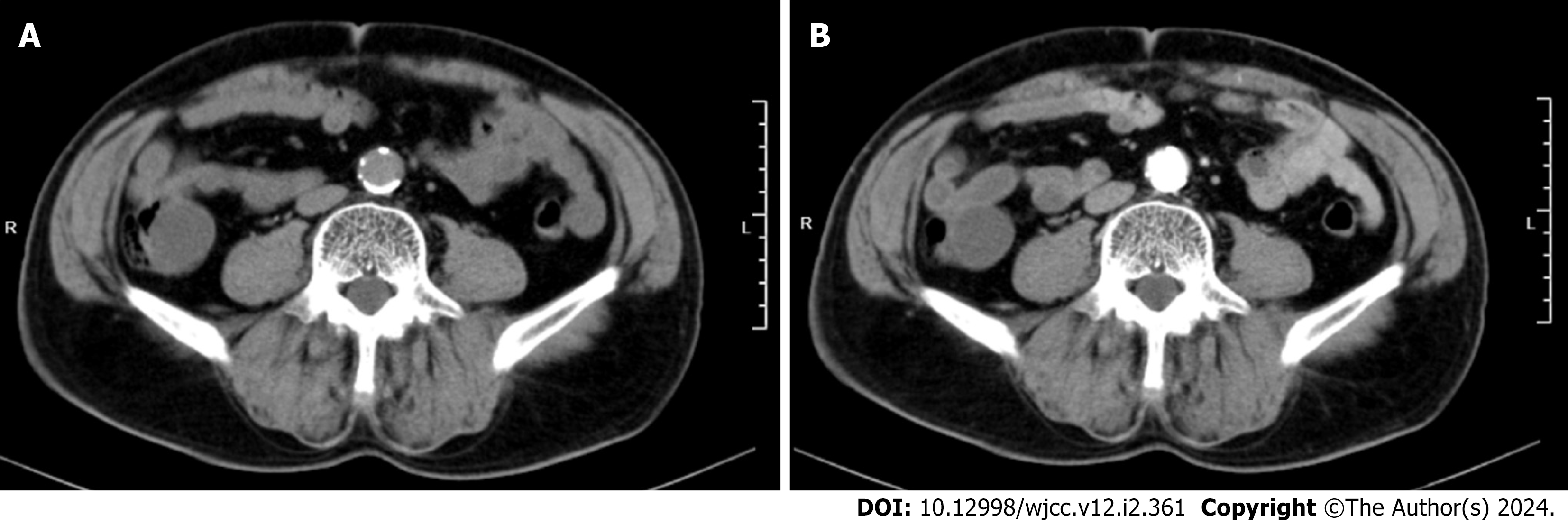
Figure 2 Abdominal computed tomography.
A: Markedly thickened and dilated appendix with visible cystic shadows; B: Appendix wall showed a slight enhancement following the contrast enhancement procedure.
Figure 3 Intraoperative view.
A: Significantly dilated appendix; B: Linear cutter/stapler was used to laparoscopically resect the appendix and portions of the cecum.
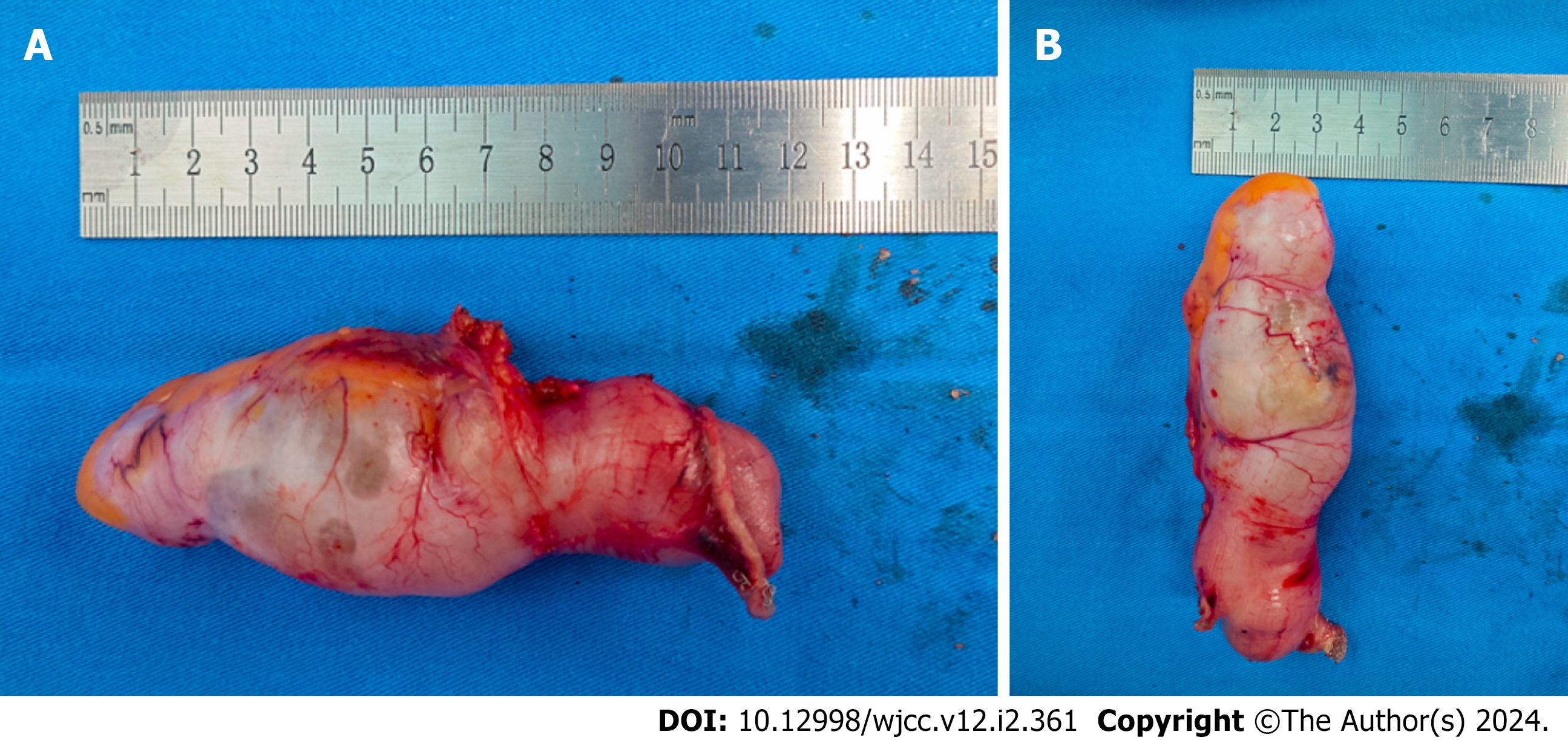
Figure 4 Appendix.
A: 12 cm in length; B: 4 cm in width.
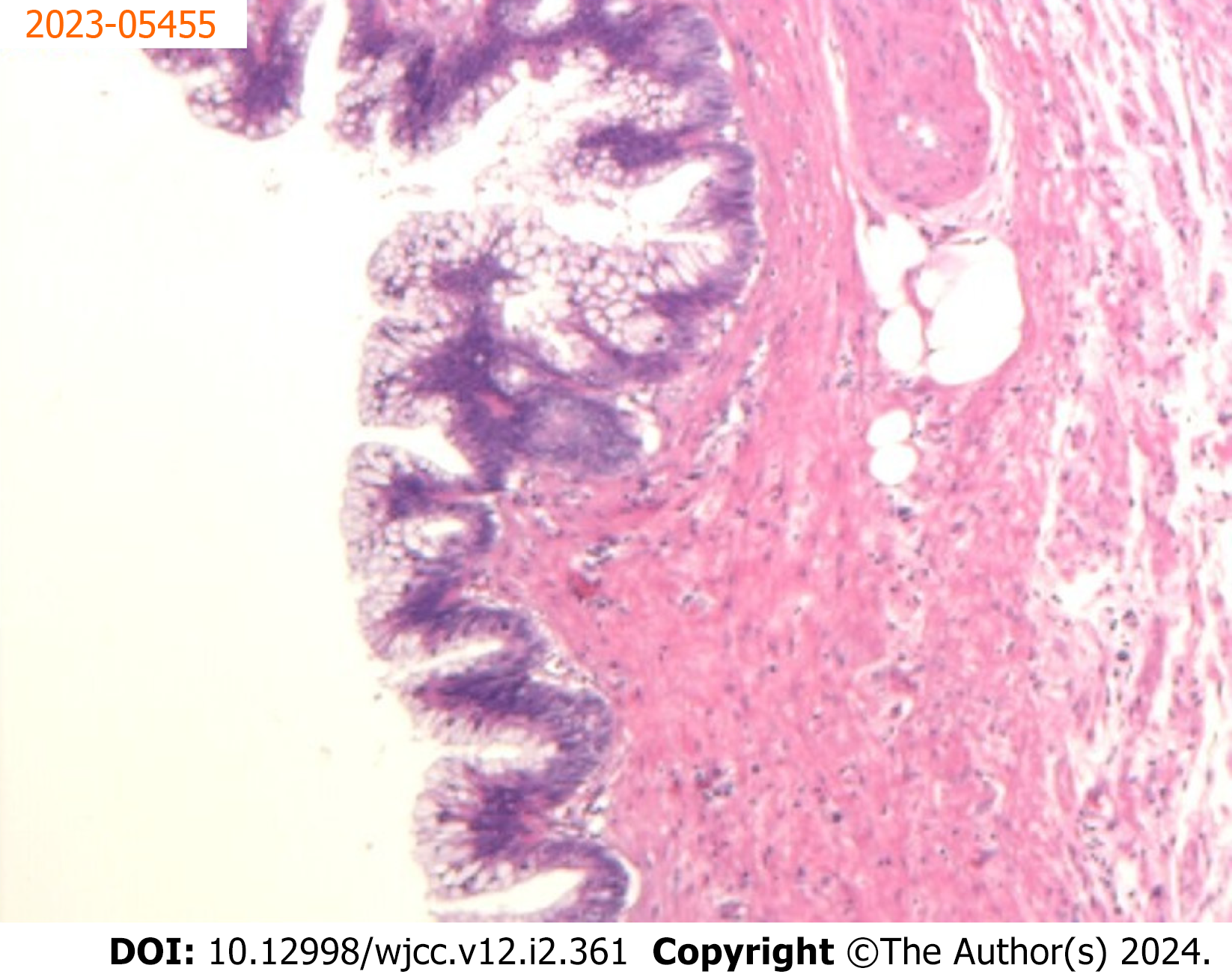
Figure 5 Postoperative pathology.
The tumor had invaded the entire appendix with mucus accumulation in the apical part of the appendix and the sub-plasma layer of the appendiceal body. The resection margins were negative (hematoxylin–eosin, × 200).
- Citation: Yao MQ, Jiang YP, Wang YY, Mou YP, Fan JX. Asymptomatic low-grade appendiceal mucinous neoplasm: A case report. World J Clin Cases 2024; 12(2): 361-366
- URL: https://www.wjgnet.com/2307-8960/full/v12/i2/361.htm
- DOI: https://dx.doi.org/10.12998/wjcc.v12.i2.361


