Copyright
©The Author(s) 2024.
World J Clin Cases. Jul 6, 2024; 12(19): 4022-4028
Published online Jul 6, 2024. doi: 10.12998/wjcc.v12.i19.4022
Published online Jul 6, 2024. doi: 10.12998/wjcc.v12.i19.4022
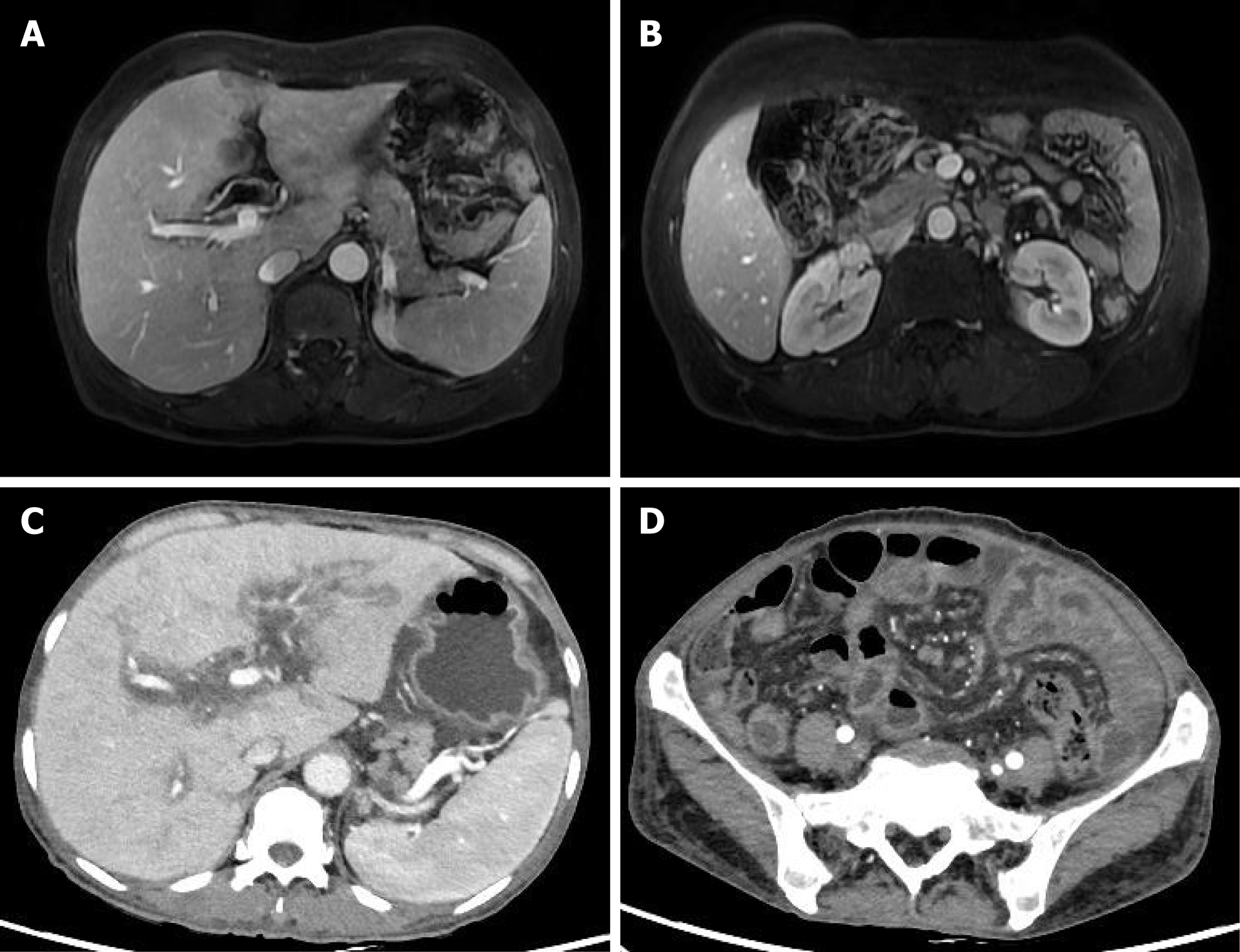
Figure 1 Abdomen magnetic resonance imaging and computed tomography findings of the female with indeterminate dendritic cell tumor.
A: Lasting contrast-enhanced of lesions in the bile duct in the magnetic resonance imaging (MRI) at the first admission; B: Enlarged mesenteric lymph nodes in MRI at the first admission; C: Tumor-like lesions in the bile duct in the computed tomography (CT) examination at the second admission; D: Enlarged mesenteric lymph nodes in the CT examination at the second admission.
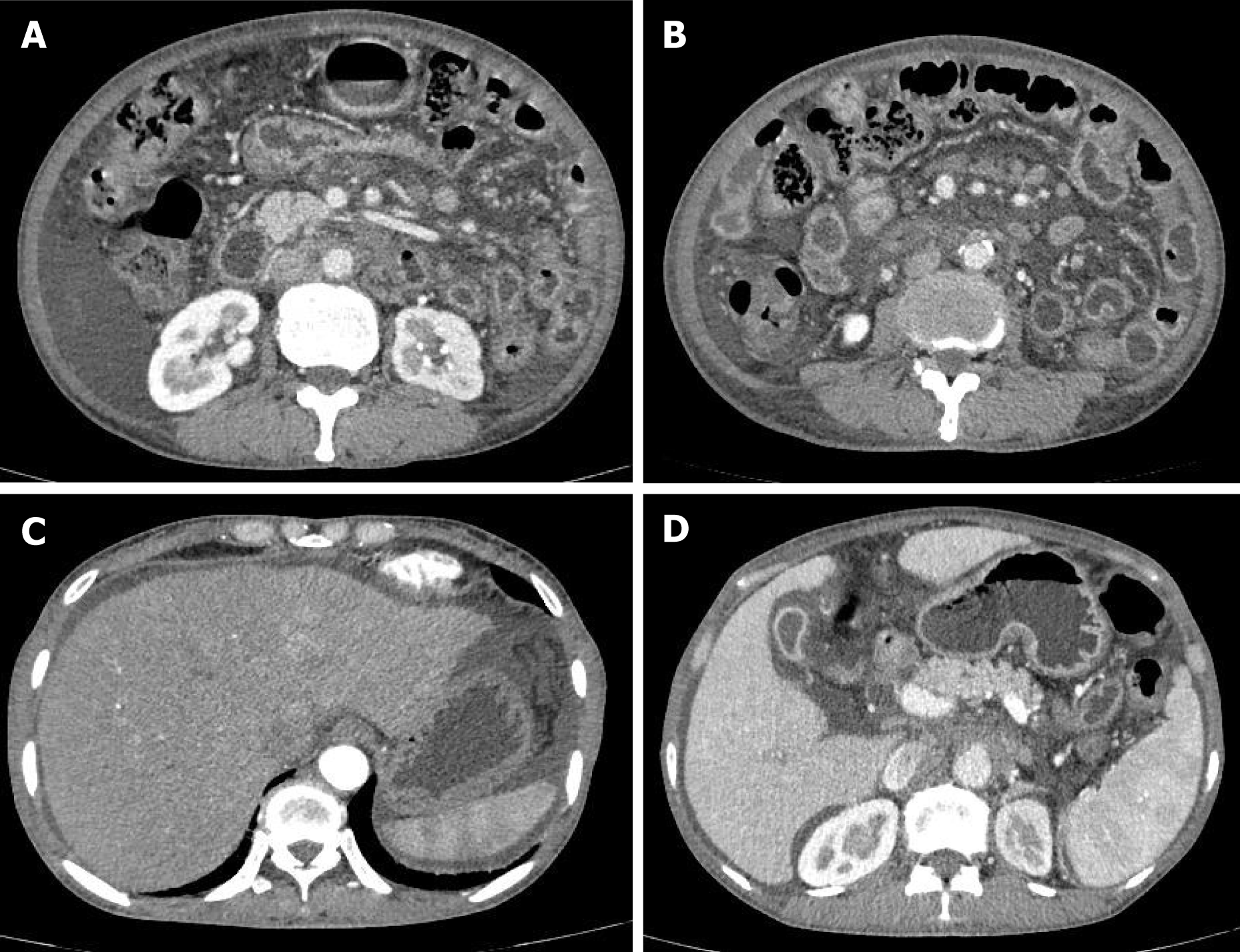
Figure 2 Increasingly enlarged mesentery lymph nodes and swollen omenta of the female with indeterminate dendritic cell tumor in the computed tomography examination.
A: Ascites and enlarged mesentery lymph nodes; B: Swollen omenta and mesentery; C: Ascites in the perihepatic areas; D: Blurred abdominal gaps.
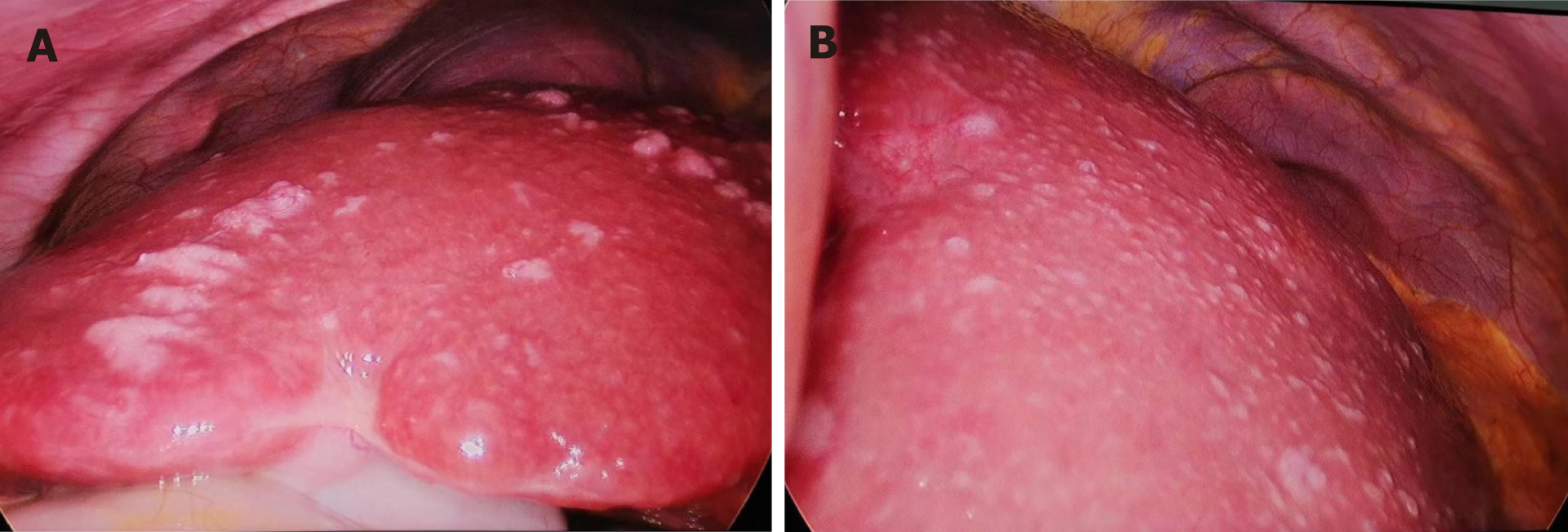
Figure 3 Diffuse lesions of Liver in the laparoscopic sights.
A: Right liver lobel; B: Left liver lobe.
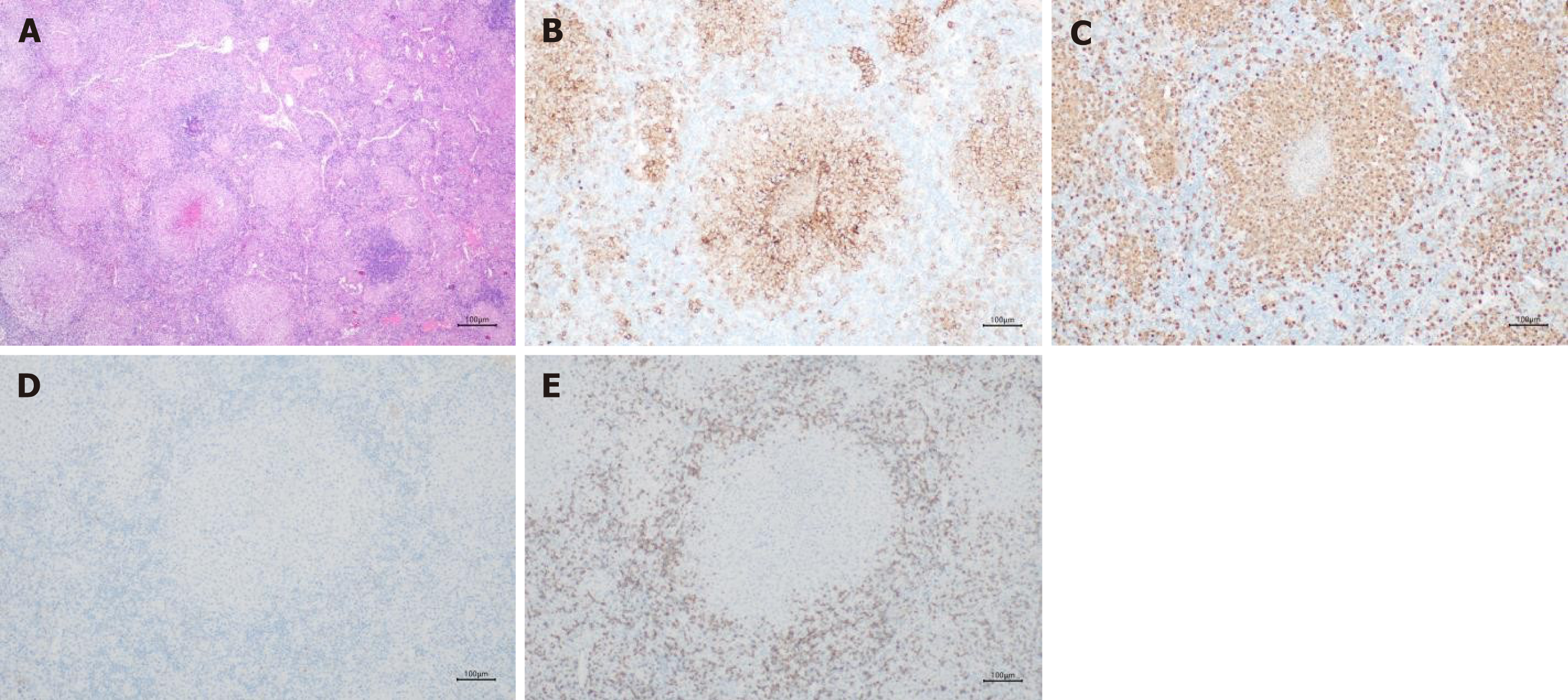
Figure 4 Histopathology and immunohistochemical analysis of indeterminate dendritic cell tumor in the lymph nodes.
A: Histopathology picture of lymph nodes (×40); B: Positive CD1a expression (×100); C: Positive s100 expression (×100); D: Negative Langerin expression (×100); E: Negative CD3 expression (×100).
- Citation: Liang H, Zhao YF, Zhang LP, Wu YK. Overall clinical course of indeterminate dendritic cell tumor patients without skin lesions: A rare case report. World J Clin Cases 2024; 12(19): 4022-4028
- URL: https://www.wjgnet.com/2307-8960/full/v12/i19/4022.htm
- DOI: https://dx.doi.org/10.12998/wjcc.v12.i19.4022


