Copyright
©The Author(s) 2024.
World J Clin Cases. Jul 6, 2024; 12(19): 3971-3977
Published online Jul 6, 2024. doi: 10.12998/wjcc.v12.i19.3971
Published online Jul 6, 2024. doi: 10.12998/wjcc.v12.i19.3971
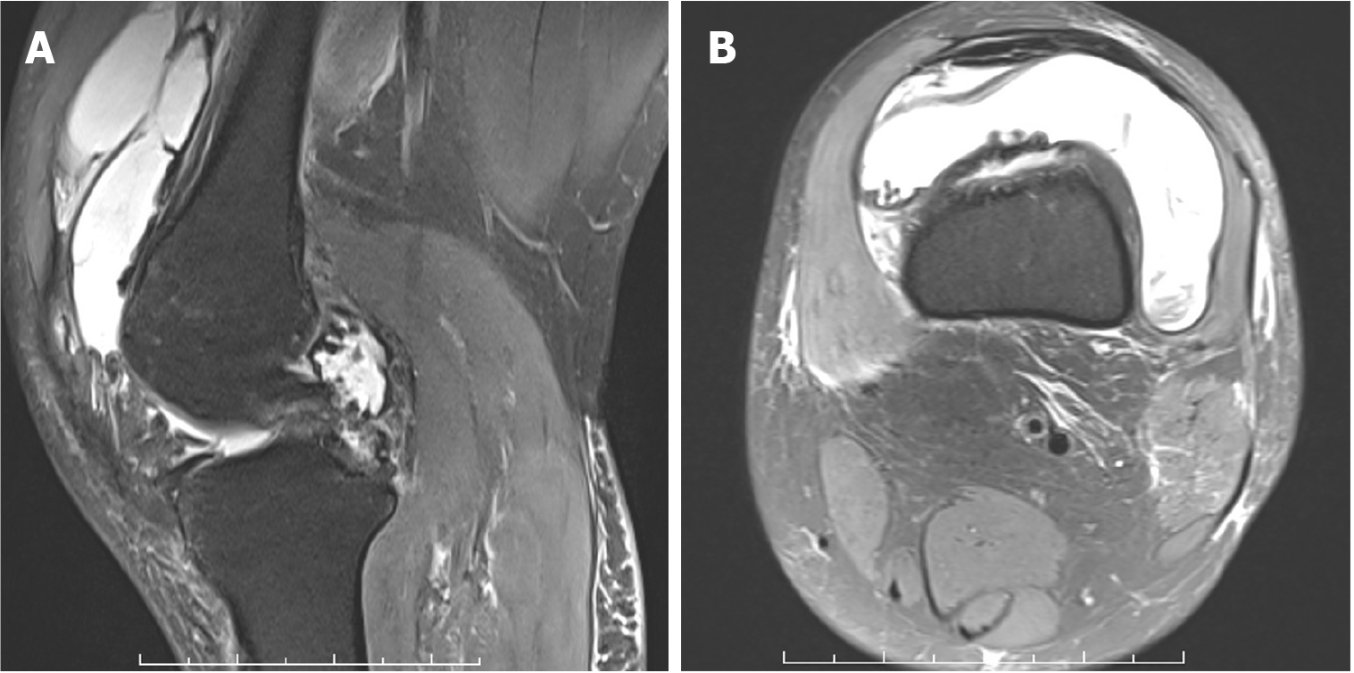
Figure 1 Magnetic resonance imagingof the left knee.
The images show joint effusion and synovial proliferation, which resemble pigmented villonodular synovitis symptoms. A: Sagittal view; B: Axial view.
- Citation: Qu YP, Jin W, Huang B, Shen J. Combination of manual lymphatic drainage and Kinesio taping for treating pigmented villonodular synovitis: A case report. World J Clin Cases 2024; 12(19): 3971-3977
- URL: https://www.wjgnet.com/2307-8960/full/v12/i19/3971.htm
- DOI: https://dx.doi.org/10.12998/wjcc.v12.i19.3971


