Copyright
©The Author(s) 2024.
World J Clin Cases. Jul 6, 2024; 12(19): 3961-3970
Published online Jul 6, 2024. doi: 10.12998/wjcc.v12.i19.3961
Published online Jul 6, 2024. doi: 10.12998/wjcc.v12.i19.3961
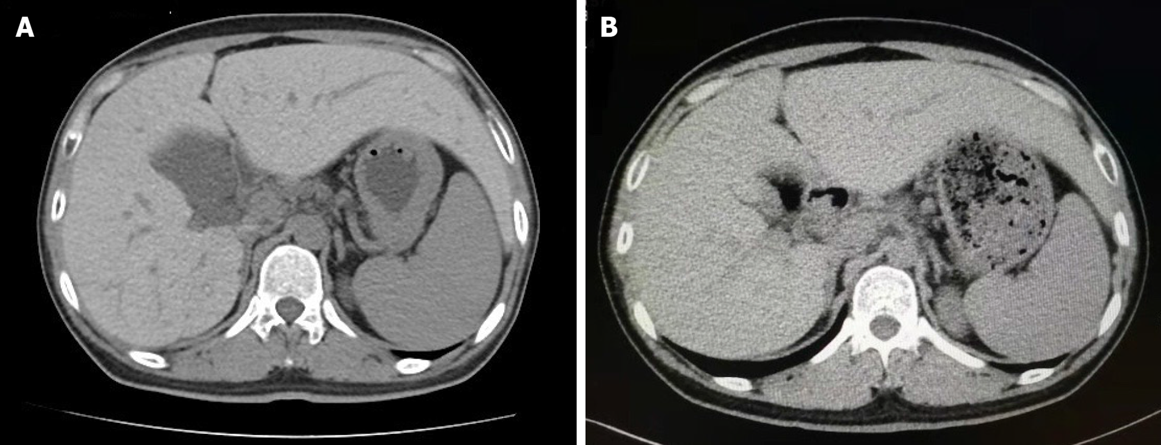
Figure 1 Computed tomography images of the abdomen.
A: Computed tomography (CT) scanning showed that the enlarged liver of the patient clearly appeared as a high-density area because of tissue iron deposition, the CT value of the liver was 104 Hounsfield unit (HU); B: The CT value of the liver decreased to 68 HU after 72 wk of phlebotomy therapy.
Figure 2 Pathological images of liver biopsy.
A: Liver biopsy sections showed preserved lobular architecture with mild to moderate portal fibrosis and a few fibrous connections; B: There was marked iron deposition (grade 4 iron stores), shown as blue granules, within hepatocytes biliary epithelial cells in the portal area and endothelial cells. Iron deposition was maximal in hepatocytes in zone 1 of the hepatic acinus and occasional in Kupffer cells.
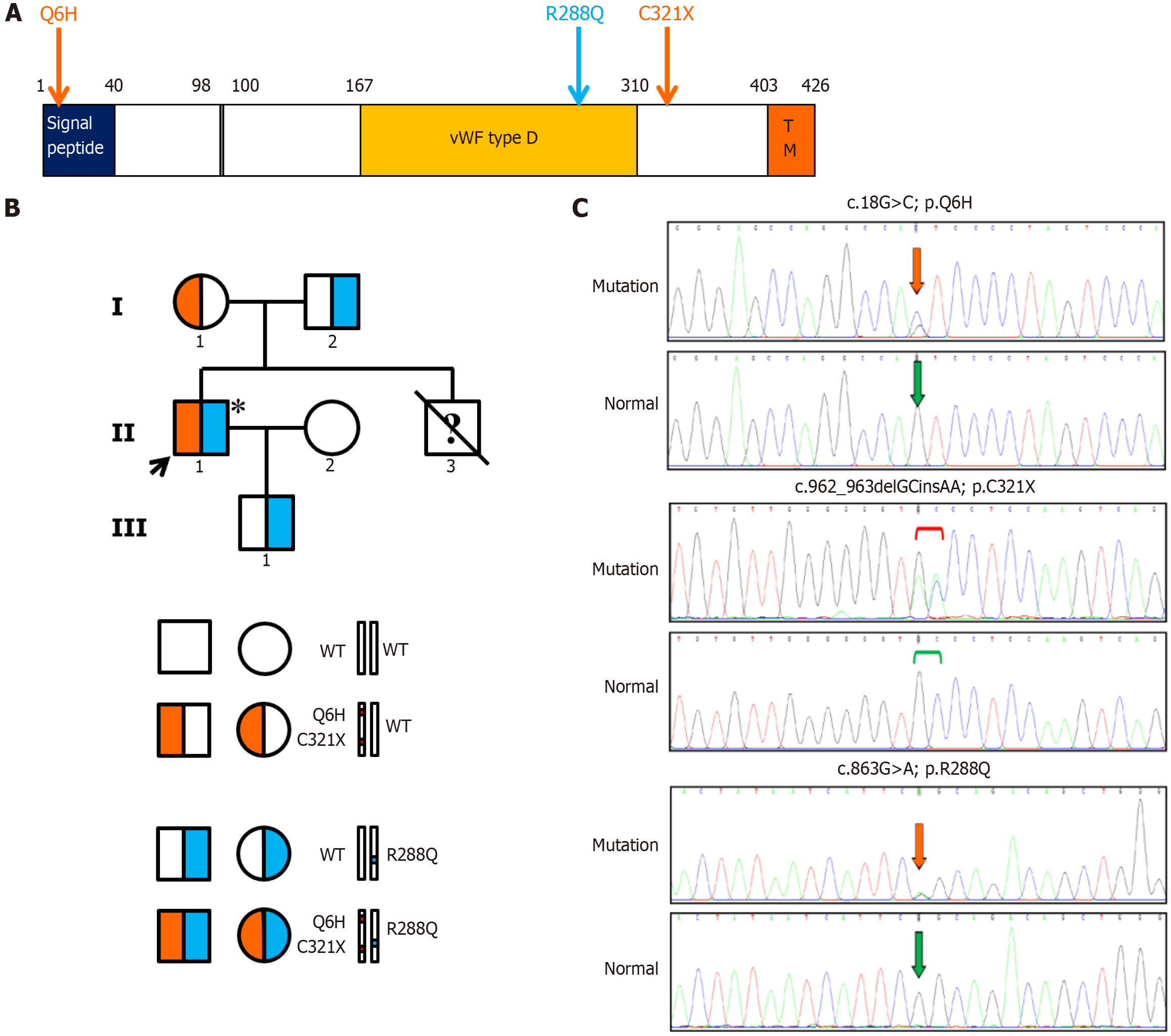
Figure 3 Hemojuvelin mutations.
A: Structure of the hemojuvelin (HJV) protein and mutations in HJV. The positions of a missense mutation Q6H and a nonsense mutation C321* which are present on the maternal allele, are marked with orange arrows. The position of a missense mutation R288Q which was found on the paternal allele, is marked with a blue arrow; B: Transmission of HJV mutations. II1 is the proband which is the only hemochromatosis patient in the family, and he carries the heterozygous mutations Q6H and C321* in cis, as well as R288Q. I1 is heterozygous for Q6H and C321* in cis; I2 and III3 are heterozygous for R288Q; II2 is wild type; the genotype of II3 is unknown. (Asterisks indicate hemochromatosis); C: Partial sequence diagram of the HJV gene. The heterozygous mutations c.18G>C, c.962_963delGCinsAA, and c.863G>A, which result in p.Q6H, p.C321*, and p.R288Q respectively, are denoted, as well as the normal sequence. vWF type D: Von willebrand factor type D domain; TM: Transmembrane domain; WT: Wide type.
- Citation: Xie LD, Kong XM, Shen JX, Wang TL, Ma J, Zhang YF, Chen XP. Novel compound heterozygous mutations in the hemojuvelin gene in a juvenile hemochromatosis patient: A case report. World J Clin Cases 2024; 12(19): 3961-3970
- URL: https://www.wjgnet.com/2307-8960/full/v12/i19/3961.htm
- DOI: https://dx.doi.org/10.12998/wjcc.v12.i19.3961


