Copyright
©The Author(s) 2024.
World J Clin Cases. Jul 6, 2024; 12(19): 3950-3955
Published online Jul 6, 2024. doi: 10.12998/wjcc.v12.i19.3950
Published online Jul 6, 2024. doi: 10.12998/wjcc.v12.i19.3950
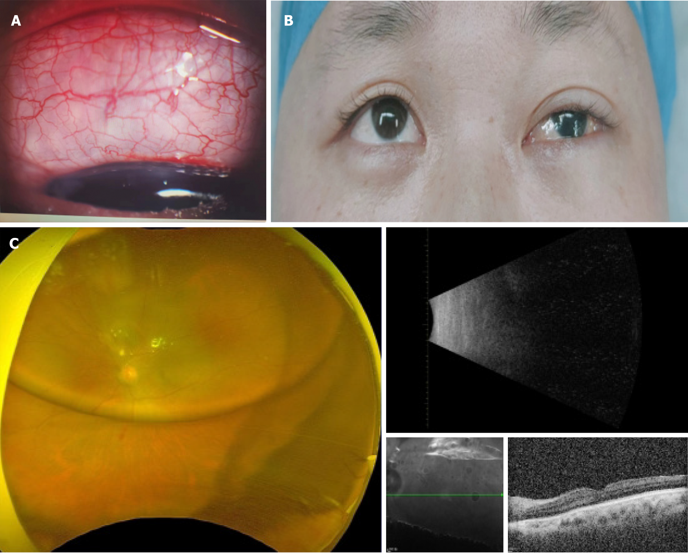
Figure 1 Preoperative ophthalmic examination.
A: After the first postoperative orbital computed tomography scan, the left eye protruded forward, with uneven thickening of the bulbar wall and elliptical high-density shadows in the eye; B: Photos of eye surface after the first operation, conjunctival foam like protrusion, oily liquid accumulation under the conjunctiva; C: Ophthalmic examination: Laser spots and silicone oil plane can be seen in fundus examination, and no obvious abnormalities are found in other examinations.
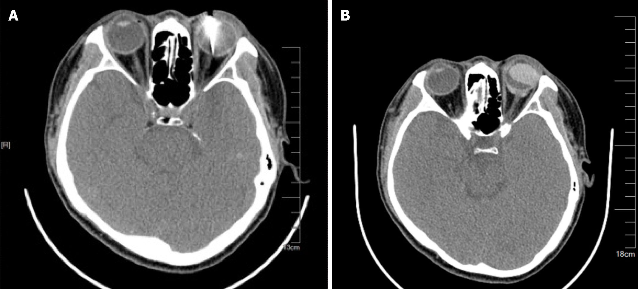
Figure 2 Preoperative orbit computed tomography scan imaging.
A: Initial diagnosis of orbital computed tomography (CT) shows high-density foreign body shadow in the left eye; B: After the first postoperative orbital CT scan, the left eye protruded forward, with uneven thickening of the bulbar wall and elliptical high-density shadows in the eye.
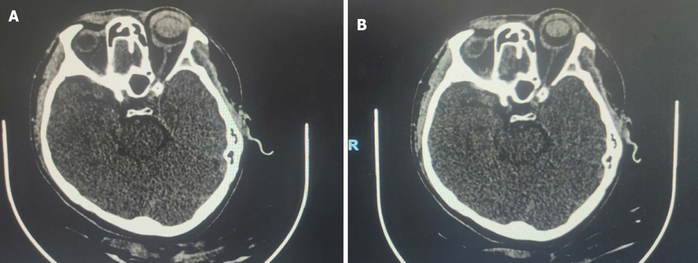
Figure 3 Postoperative follow-up comparison.
Comparison of orbital computed tomography before and after the second surgery shows a significant reduction in the migration of silicone oil. A: Before; B: After.
- Citation: Shu BL, Wu HY, Hu YX, Rao J, Wei B, Huang QY, Wu XR. Silicone oil migrating into the conjunctival space and orbit after surgery for an eye-penetrating injury: A case report. World J Clin Cases 2024; 12(19): 3950-3955
- URL: https://www.wjgnet.com/2307-8960/full/v12/i19/3950.htm
- DOI: https://dx.doi.org/10.12998/wjcc.v12.i19.3950


