Copyright
©The Author(s) 2024.
World J Clin Cases. Jul 6, 2024; 12(19): 3931-3935
Published online Jul 6, 2024. doi: 10.12998/wjcc.v12.i19.3931
Published online Jul 6, 2024. doi: 10.12998/wjcc.v12.i19.3931
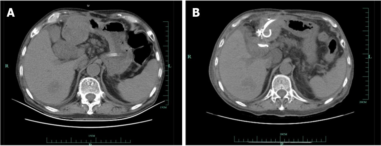
Figure 1 Computed tomography showed the abdominal abscess before and after the drainage.
A: Computed tomography (CT) showed that a big low-density dumbbell-shaped mass among the liver and intestine; B: One day after operation, CT showed that the low-density mass was reduced.
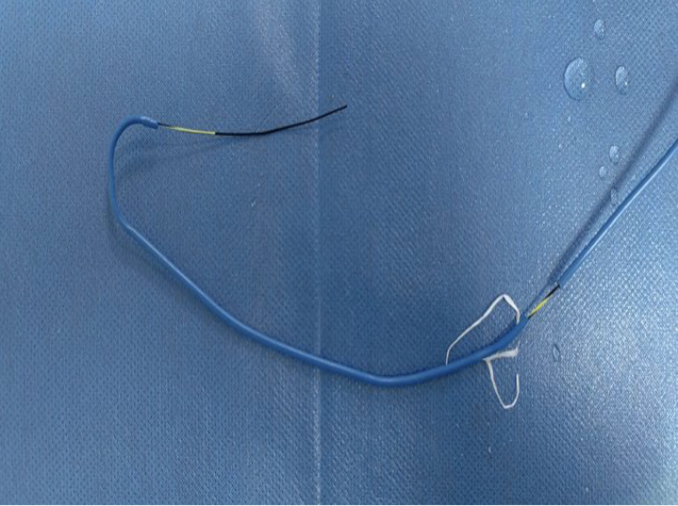
Figure 2 The special stent device.
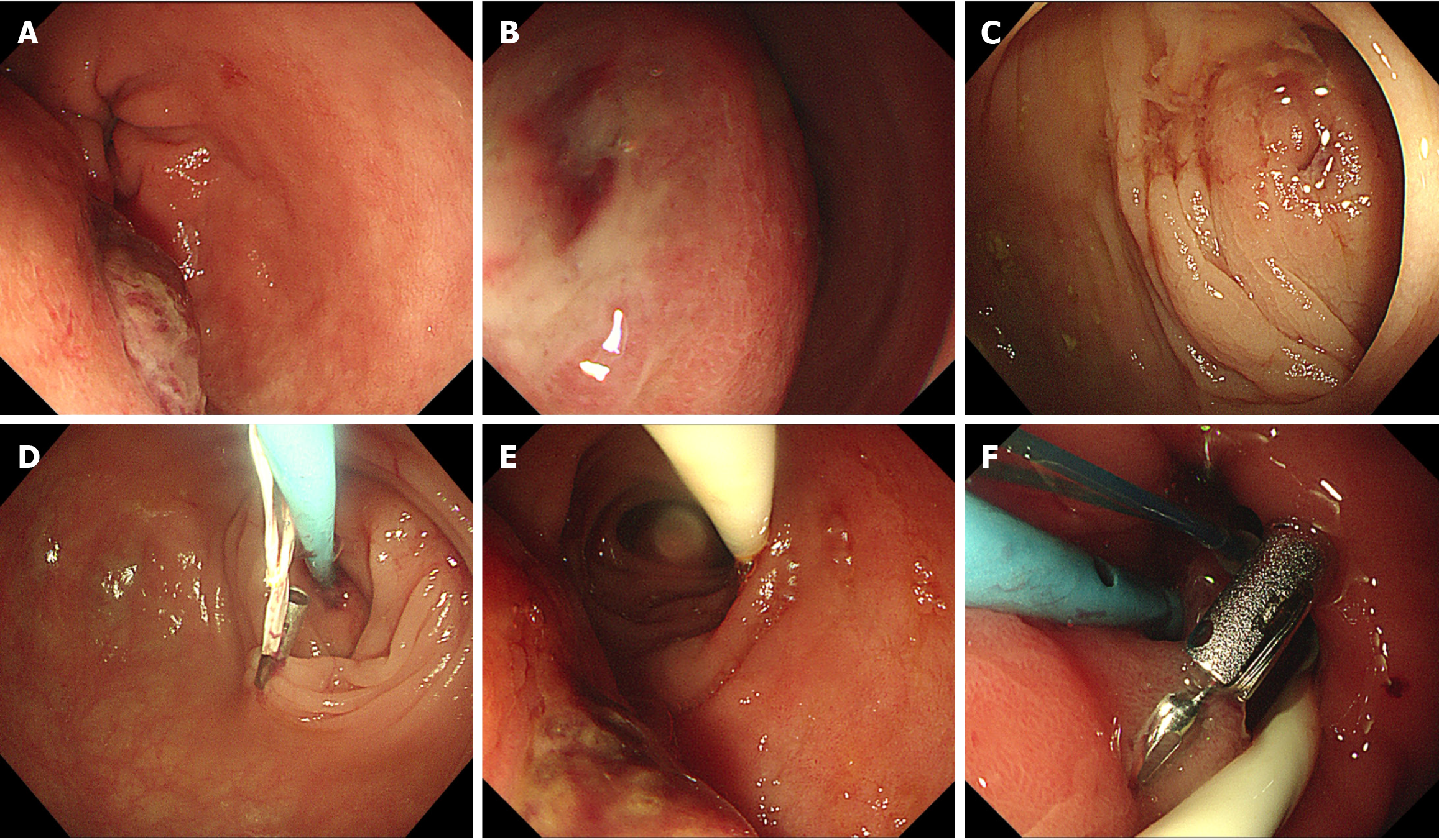
Figure 3 Special stent for draining the abdominal abscess respectively from colon and duodenum under endoscopy.
A: Gastroscopy showed a big rupture on the submucosal mass at the descending duodenum; B: Gastroscopy showed a fistula on the submucosal mass at the duodenal bulb; C: Colonoscopy showed a submucosal mass with a fistula at the colon of liver region; D: The stent was placed into the mass through the fistula; E: A nasojejunal nutrient tube was retained to the jejunum; F: The special stent used to drain the mass from the duodenal bulb.
- Citation: Zhang FL, Xu J, Jiang YH, Zhu YD, Wu QN, Shi Y, Zhan ZY, Wang H. Special stent for draining the abdominal abscess respectively from colon and duodenum: A case report. World J Clin Cases 2024; 12(19): 3931-3935
- URL: https://www.wjgnet.com/2307-8960/full/v12/i19/3931.htm
- DOI: https://dx.doi.org/10.12998/wjcc.v12.i19.3931


