Copyright
©The Author(s) 2024.
World J Clin Cases. Jul 6, 2024; 12(19): 3918-3924
Published online Jul 6, 2024. doi: 10.12998/wjcc.v12.i19.3918
Published online Jul 6, 2024. doi: 10.12998/wjcc.v12.i19.3918
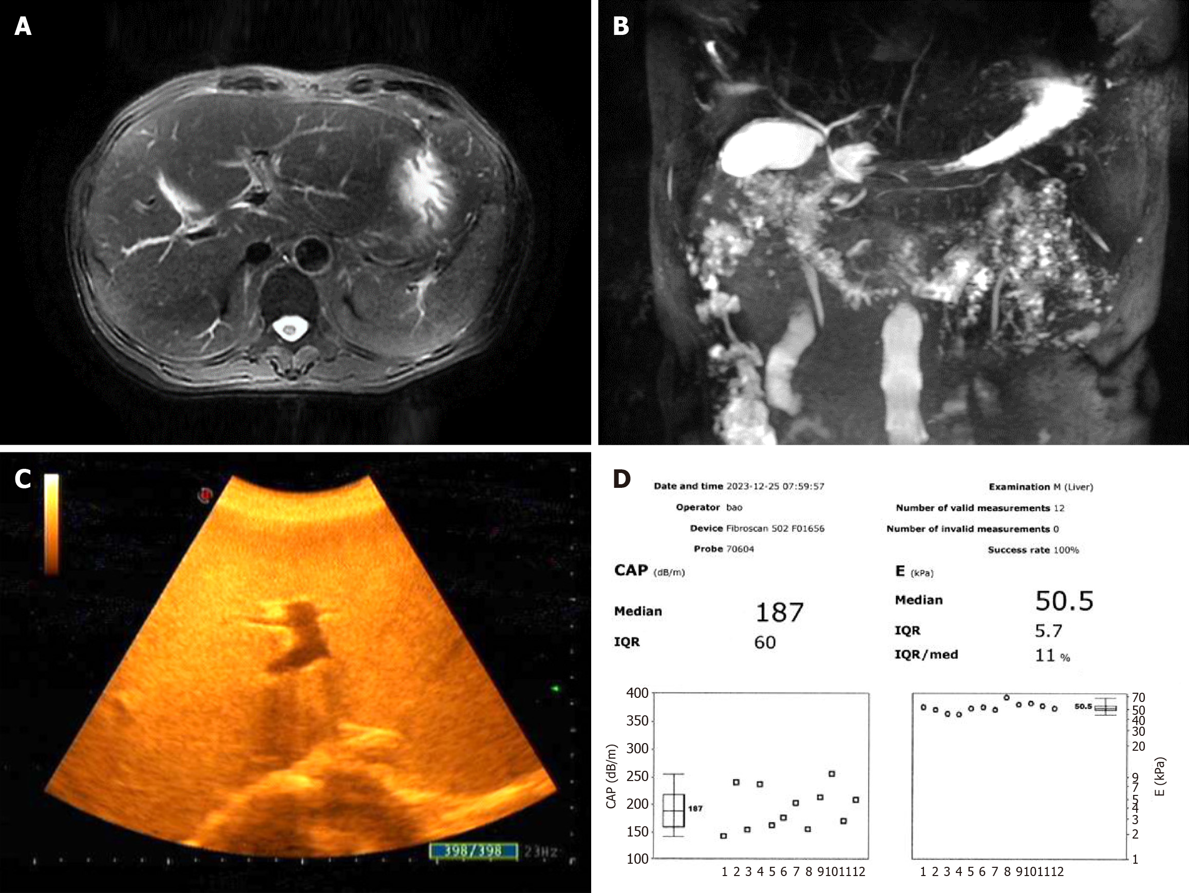
Figure 1 Imaging examinations.
A: The image of abdominal magnetic resonance imaging; B: The image of magnetic resonance cholangiopancreatography; C: The image of abdominal B-mode ultrasound; D: The results of FibroScan. IQR: Interquartile range.
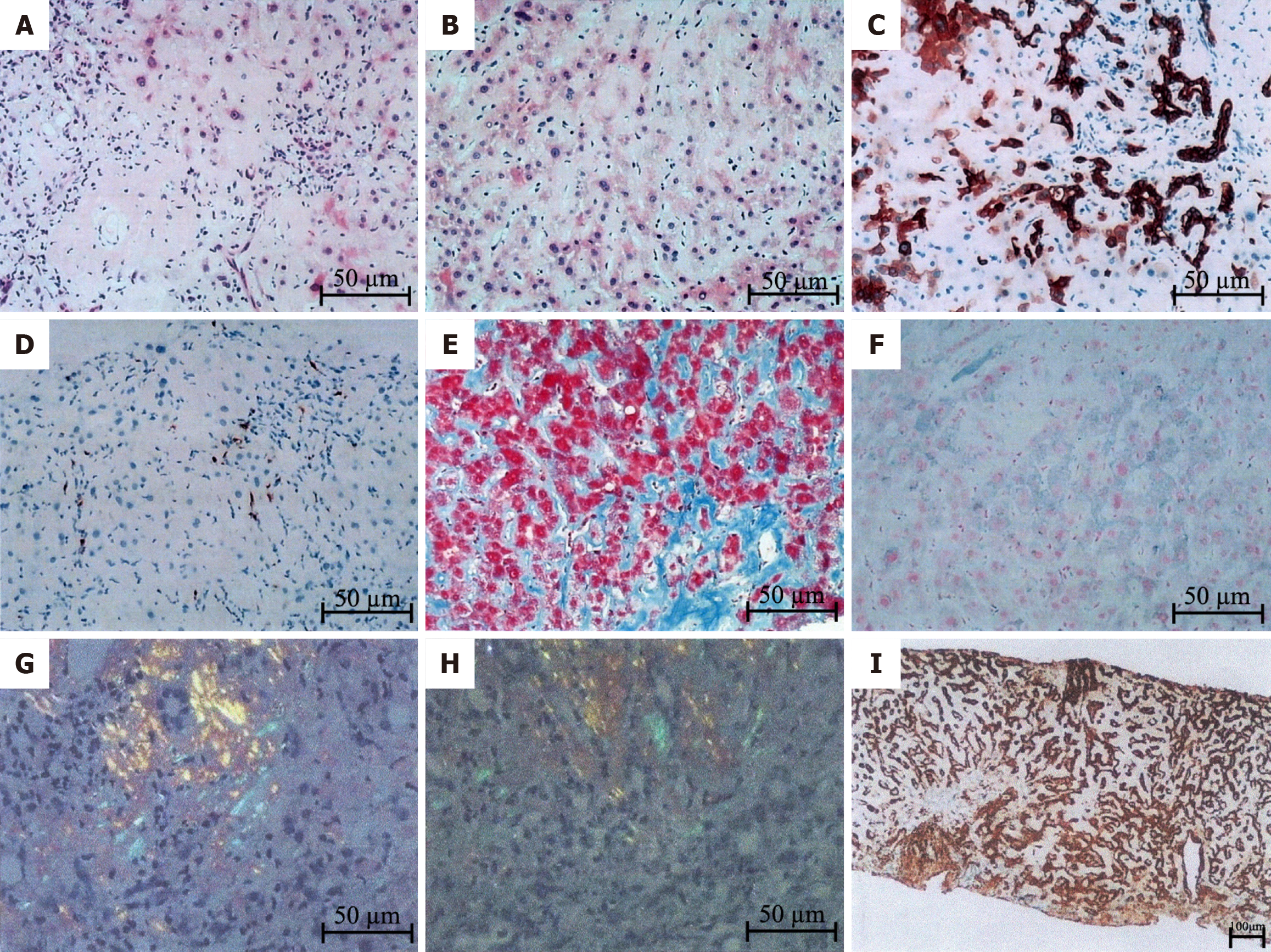
Figure 2 Liver biopsy and pathological examination.
A: Diffuse deposition of pale pink homogeneous material was observed in the perisinusoidal space, hepatocyte space, and subendothelial space of the central vein; B: Deposition of homogeneous pale pink material in perisinus space and hepatocellular atrophy were observed; C: Positive CK7 staining in the bile duct epithelium and in the approximately 90% of liver cells were observed; D: Positive MUM1 staining in a few plasma cells; E: Masson staining revealed diffuse deposition of homogeneous material around the sinusoids and blood vessels; F: Prussian blue staining showed the deposition of iron particles in liver cells and Kupffer cells; G: Polarized light microscopy revealed positive Congo red staining; H: Polarized light microscopy showed positive oxidized Congo red staining; I: Immunohistochemical results showed positive Ig κ staining.
- Citation: Chen Y, Peng J, Wang Y, Xiao LH, Liu F, Wei YB, Wu XF, Wang LW. Hepatic amyloidosis in a patient with chronic liver failure: A case report. World J Clin Cases 2024; 12(19): 3918-3924
- URL: https://www.wjgnet.com/2307-8960/full/v12/i19/3918.htm
- DOI: https://dx.doi.org/10.12998/wjcc.v12.i19.3918


