Copyright
©The Author(s) 2024.
World J Clin Cases. Jun 26, 2024; 12(18): 3636-3643
Published online Jun 26, 2024. doi: 10.12998/wjcc.v12.i18.3636
Published online Jun 26, 2024. doi: 10.12998/wjcc.v12.i18.3636
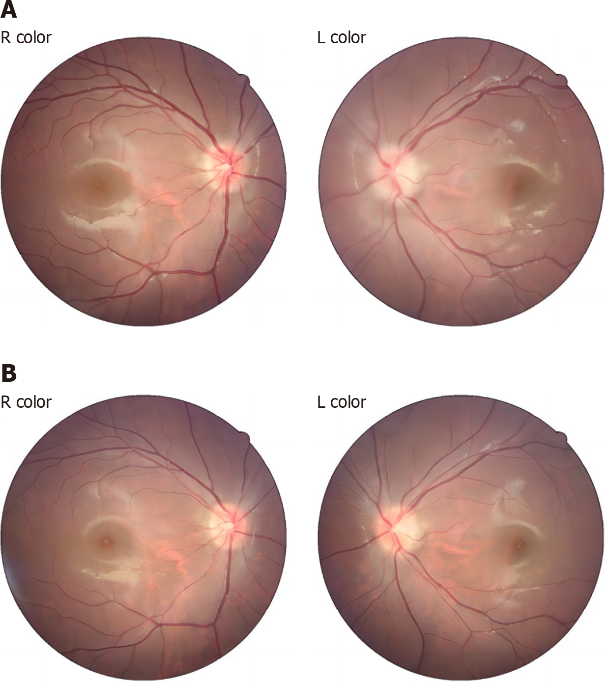
Figure 1 Fundus photographic examination results.
A: Pretreatment; B: Posttreatment.
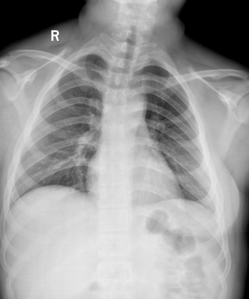
Figure 2
Analysis of chest X-ray results.
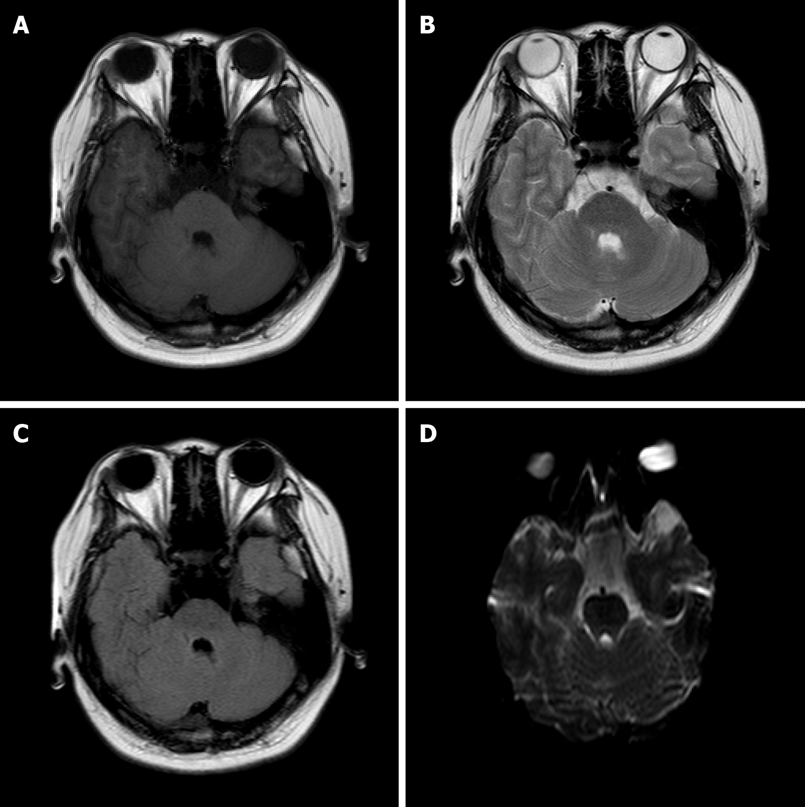
Figure 3 Results of craniocerebral magnetic examination.
A: T1-weighted imaging; B: T2-weighted imaging; C: Fluid-attenuated inversion recovery; D: Diffusion-weighted imaging.
Figure 4 Ocular B ultrasonography results.
A: Left eye; B: Right eye.
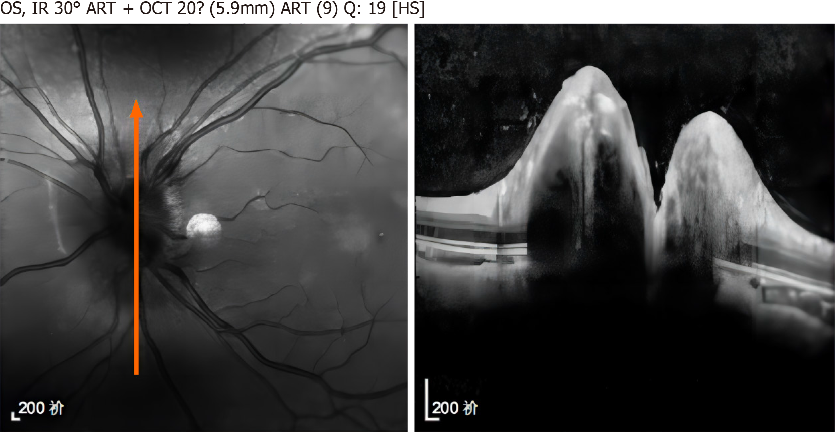
Figure 5
Optical coherence tomography examination.
- Citation: Zhen YY, Yang J, Liao PY. Human herpesvirus 7 meningitis in an adolescent with normal immune function: A case report. World J Clin Cases 2024; 12(18): 3636-3643
- URL: https://www.wjgnet.com/2307-8960/full/v12/i18/3636.htm
- DOI: https://dx.doi.org/10.12998/wjcc.v12.i18.3636


