Copyright
©The Author(s) 2024.
World J Clin Cases. Jun 26, 2024; 12(18): 3622-3628
Published online Jun 26, 2024. doi: 10.12998/wjcc.v12.i18.3622
Published online Jun 26, 2024. doi: 10.12998/wjcc.v12.i18.3622
Figure 1 Abdominopelvic computed tomography findings.
An eccentric broad-based delayed-enhancing mass-like lesion in medial wall of the second duodenum (arrow).
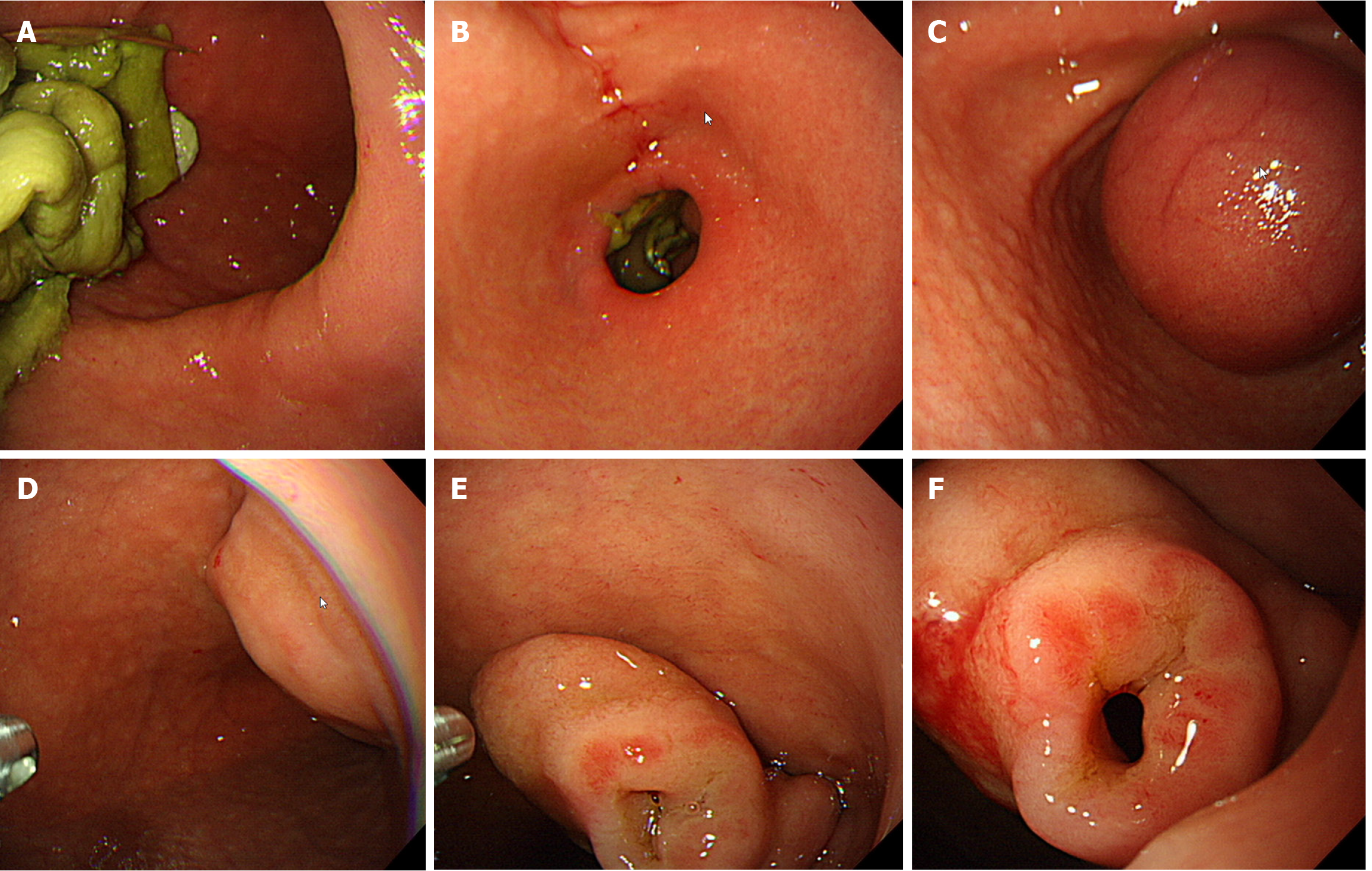
Figure 2 Initial endoscopic findings.
A: Despite a long period of fasting, large amounts of vegetable food residues are observed in the gastric cavity; B: Beyond the pylorus, food residues are retained in the duodenal bulb; C: The lesion is invaginated proximally and observed as a subepithelial tumor in the gastric cavity; D: The lumen of the duodenal bulb is significantly dilated; E and F: A hole is observed eccentrically in the elongated web.
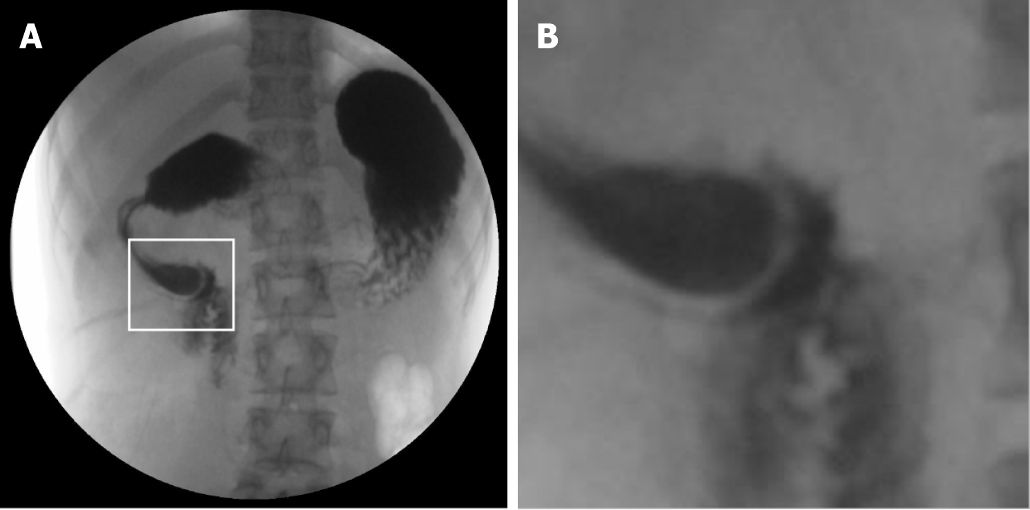
Figure 3 Gastrografin upper gastrointestinal series.
A: Intraduodenal barium contrast-filled sac with a curvilinear narrow radiolucent rim; B: An enlarged version of the photograph inside the square in panel A, showing a typical "windsock" sign.
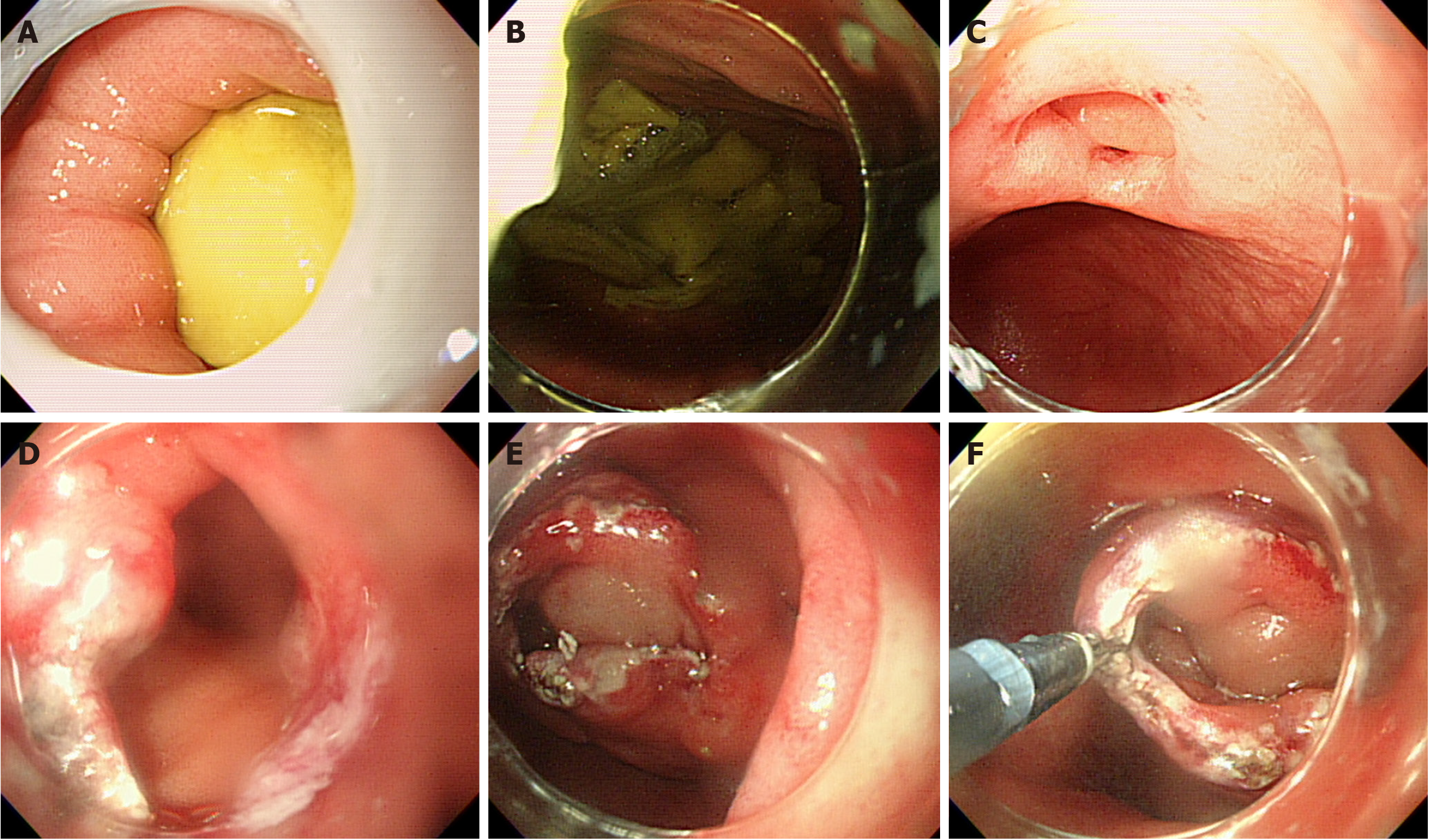
Figure 4 Endoscopic incision and cutting procedure.
A: Food residues retained in the duodenal bulb through the pylorus are observed; B: Retained food residues were expelled into the gastric cavity; C: The web is projected distally, so that the end appears like a blind pouch, and the aperture is observed at 12 o'clock; D: After attaching a transparent cap to the tip of the endoscope, a small radial incision was carefully made into the web using a IT2-knife; E: In the center, toward the duodenal wall, a long radial incision was made; F: The web was cut circumferentially along the duodenal wall.
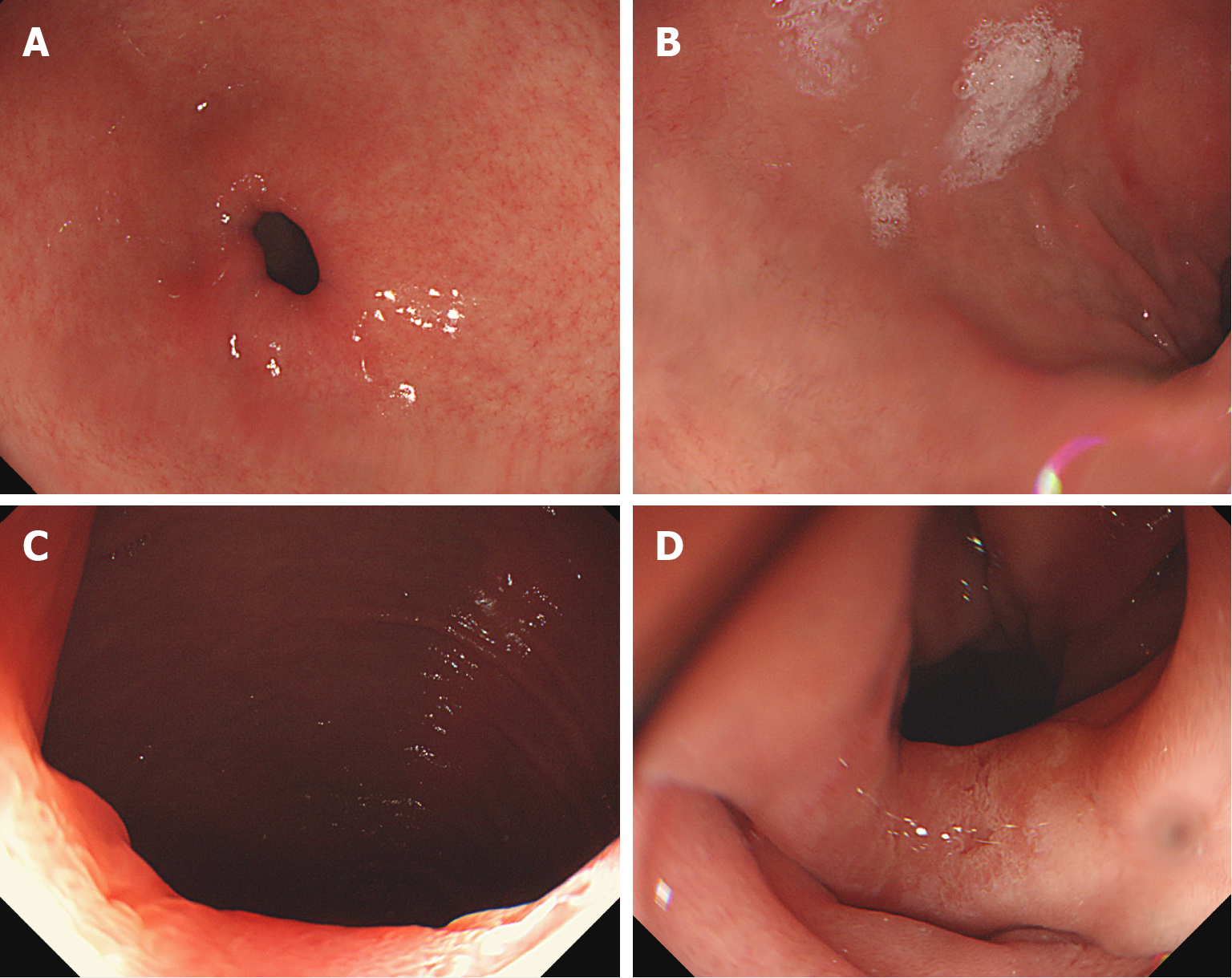
Figure 5 Last follow-up of endoscopy.
A: As in previous tests, food retention is not observed; B: The duodenal bulb is also not significantly dilated as in the previous examinations; C and D: A portion of the residual web is observed at 6 o’clock, although a sufficient width of the lumen is secured.
- Citation: Shin HD. Endoscopic radial incision and cutting method for adult congenital duodenal webs: A case report. World J Clin Cases 2024; 12(18): 3622-3628
- URL: https://www.wjgnet.com/2307-8960/full/v12/i18/3622.htm
- DOI: https://dx.doi.org/10.12998/wjcc.v12.i18.3622


