Copyright
©The Author(s) 2024.
World J Clin Cases. Jun 26, 2024; 12(18): 3603-3608
Published online Jun 26, 2024. doi: 10.12998/wjcc.v12.i18.3603
Published online Jun 26, 2024. doi: 10.12998/wjcc.v12.i18.3603
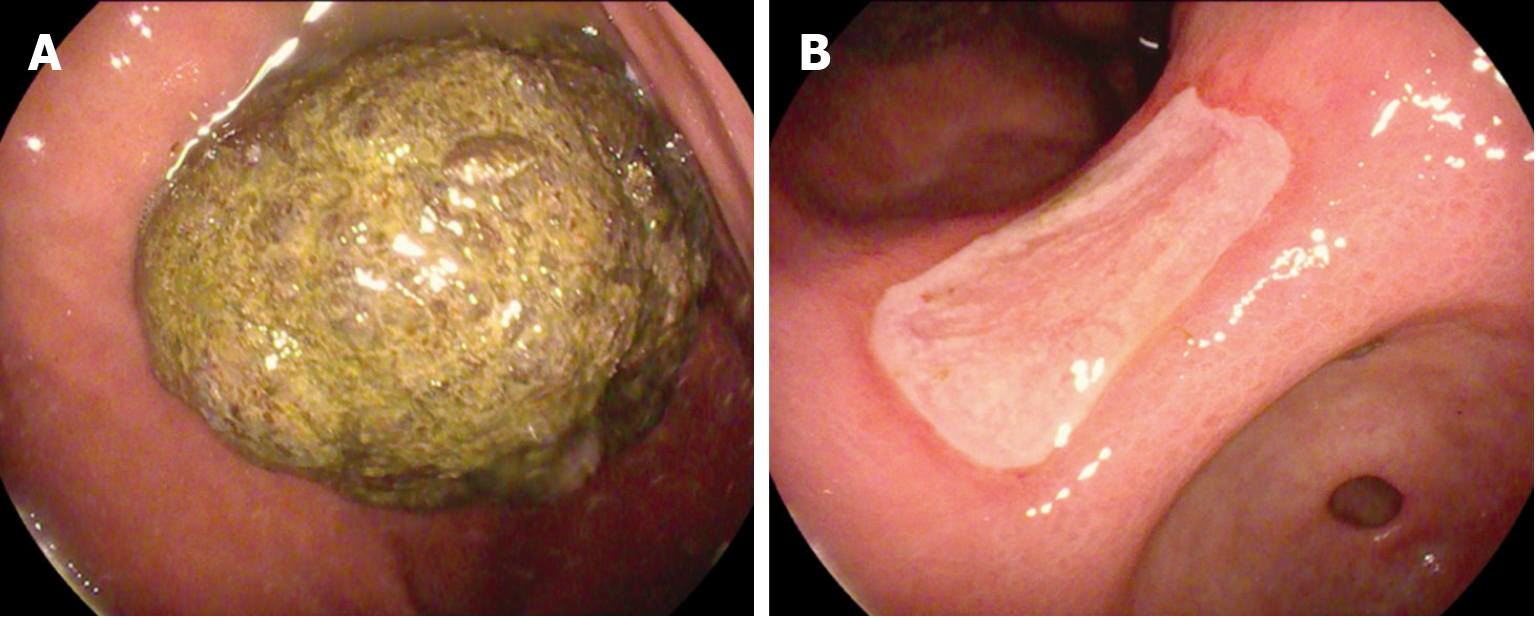
Figure 1 Initial gastroscopy images.
A: A large phytobezoar in the gastric lumen; B: A superficial ulcer in the stomach.
Figure 2 Equipment used during surgery.
A: A pre-sterilized tennis ball cord; B: A freely adjustable coil consisting of a tennis ball cord and an endoscope; C: An assistant helping to fix the tennis ball cord outside the mirror body.
Figure 3 Treatment of the gastric phytobezoar.
A: A huge gastric phytobezoar strangulated with the tennis ball cord; B: A gastric phytobezoar less than 2 cm in diameter strangulated with a trap; C: A tiny gastric phytobezoar after lithotripsy, which can be excreted through the intestine.
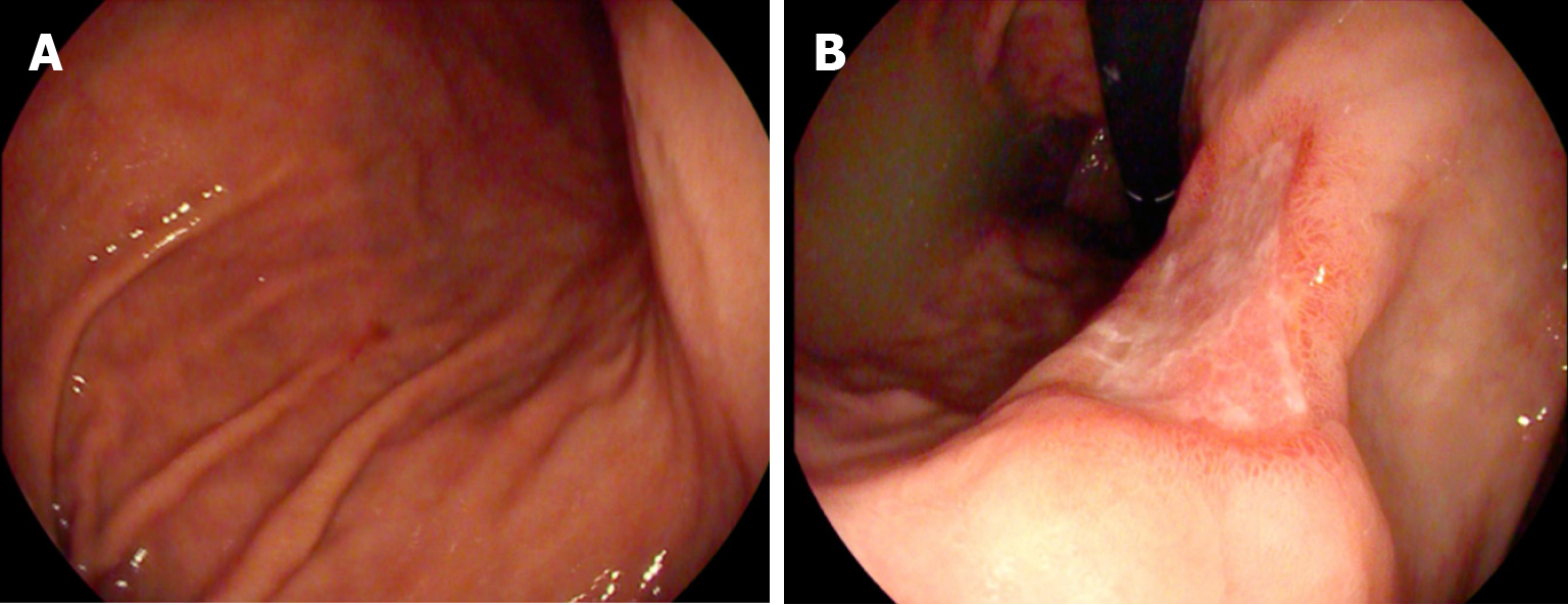
Figure 4 Gastroscopy was repeated 3 d after lithotripsy.
A: All the gastric phytobezoars had been expelled; B: Healing of the gastric ulcer.
- Citation: Shu J, Zhang H. Tennis ball cord combined with endoscopy for giant gastric phytobezoar: A case report. World J Clin Cases 2024; 12(18): 3603-3608
- URL: https://www.wjgnet.com/2307-8960/full/v12/i18/3603.htm
- DOI: https://dx.doi.org/10.12998/wjcc.v12.i18.3603


