Copyright
©The Author(s) 2024.
World J Clin Cases. Jun 26, 2024; 12(18): 3575-3581
Published online Jun 26, 2024. doi: 10.12998/wjcc.v12.i18.3575
Published online Jun 26, 2024. doi: 10.12998/wjcc.v12.i18.3575
Figure 1 The distal of the mandibular left second molar presented the non-keratinized mucosa.
A: The view from the buccal facet; B: The view from the lingual facet; C: The view from the occlusal facet.
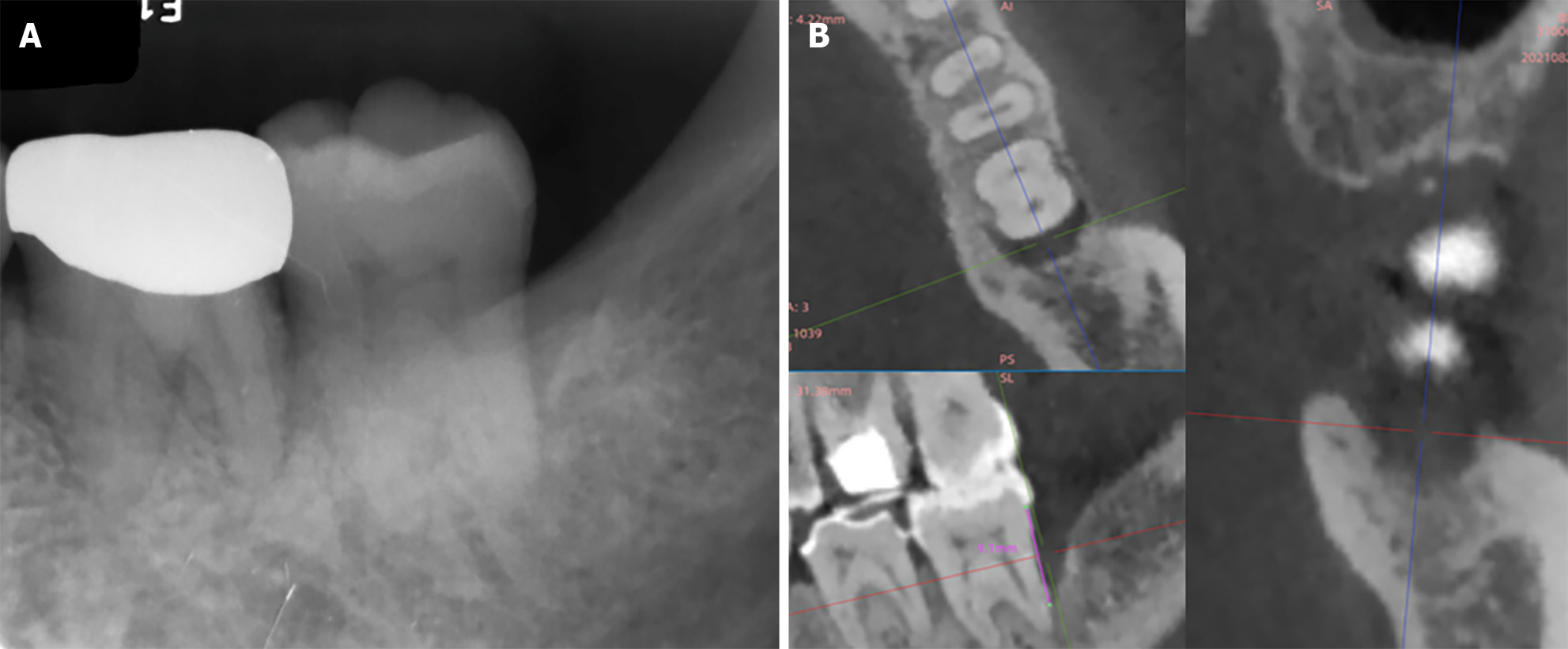
Figure 2 The preoperative radiographic images of the alveolar bone around the mandibular left second molars.
A: The parallel film demonstrated an angular bone defect at the distal of the second molar; B: Three-wall intrabony defect was found at this site on cone-beam computed tomography image.
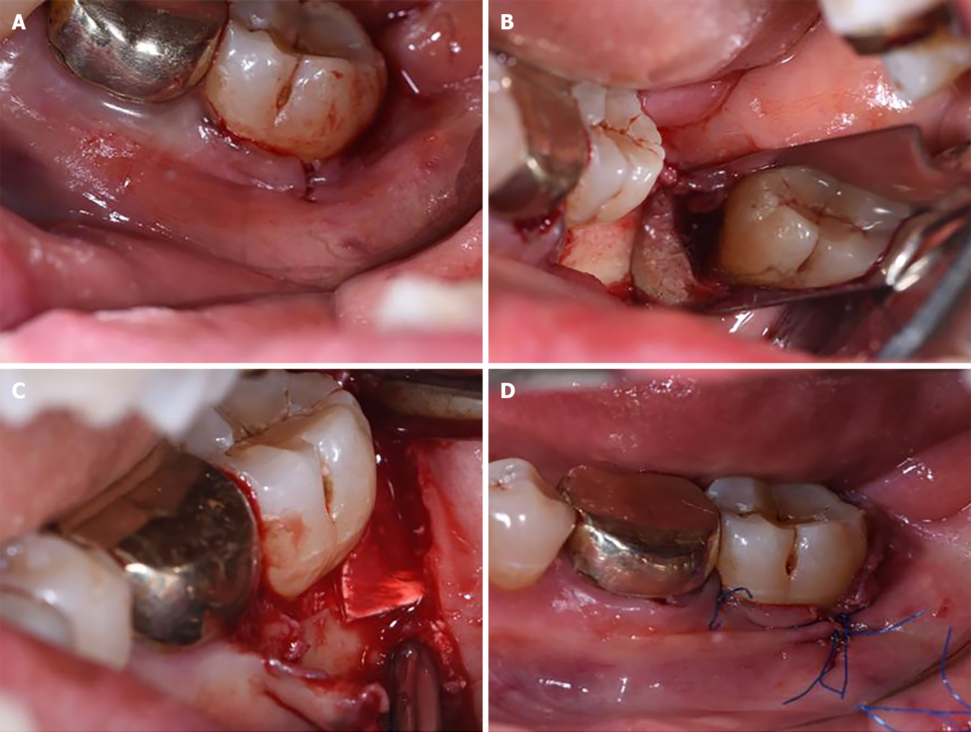
Figure 3 The modified procedure of the regenerative surgery on the mandibular left second molar.
A: The vertical incision was made on the distobuccal keratinized gingiva of the second molar; B: The whole distal non-keratinized soft tissue was tunnel-like elevated, and a 3 wall-intrabony defect was exposed; C: The intrabony defects was filled with deproteinized bovine bone mineral and covered with collagen membrane; D: The flap was positioned and sutured tightly. The vertical incision was closed with interrupted and anchored suture.
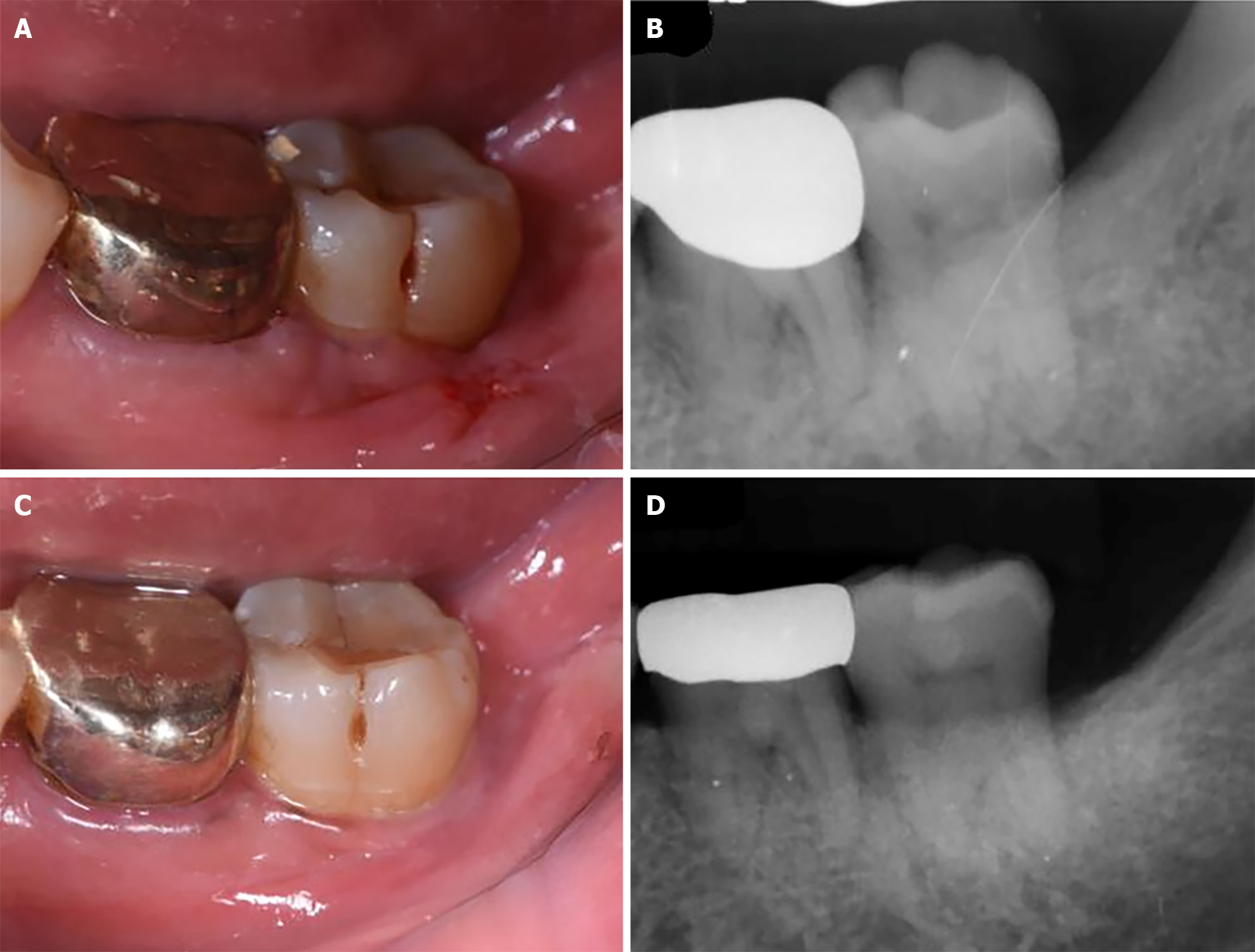
Figure 4 The outcomes of regenerative treatment for the mandibular left second molars.
A: Primary wound healing at the 2 wk after surgery; B: The parallel film showed 100% bone fill in the distal intrabony defects at 2 wk postoperatively; C: No gingiva accession appeared around the second molar at 9 months postoperatively; D: The radiographic bone gain remained satisfactory at the distal site according to the parallel film.
- Citation: Liu JR, Huang Y, Ouyang XY, Liu WY, Xie Y. Modified approach of regenerative treatment for distal intrabony defect beneath non-keratinized mucosa at terminal molar: A case report. World J Clin Cases 2024; 12(18): 3575-3581
- URL: https://www.wjgnet.com/2307-8960/full/v12/i18/3575.htm
- DOI: https://dx.doi.org/10.12998/wjcc.v12.i18.3575


