Copyright
©The Author(s) 2024.
World J Clin Cases. Jun 26, 2024; 12(18): 3534-3538
Published online Jun 26, 2024. doi: 10.12998/wjcc.v12.i18.3534
Published online Jun 26, 2024. doi: 10.12998/wjcc.v12.i18.3534
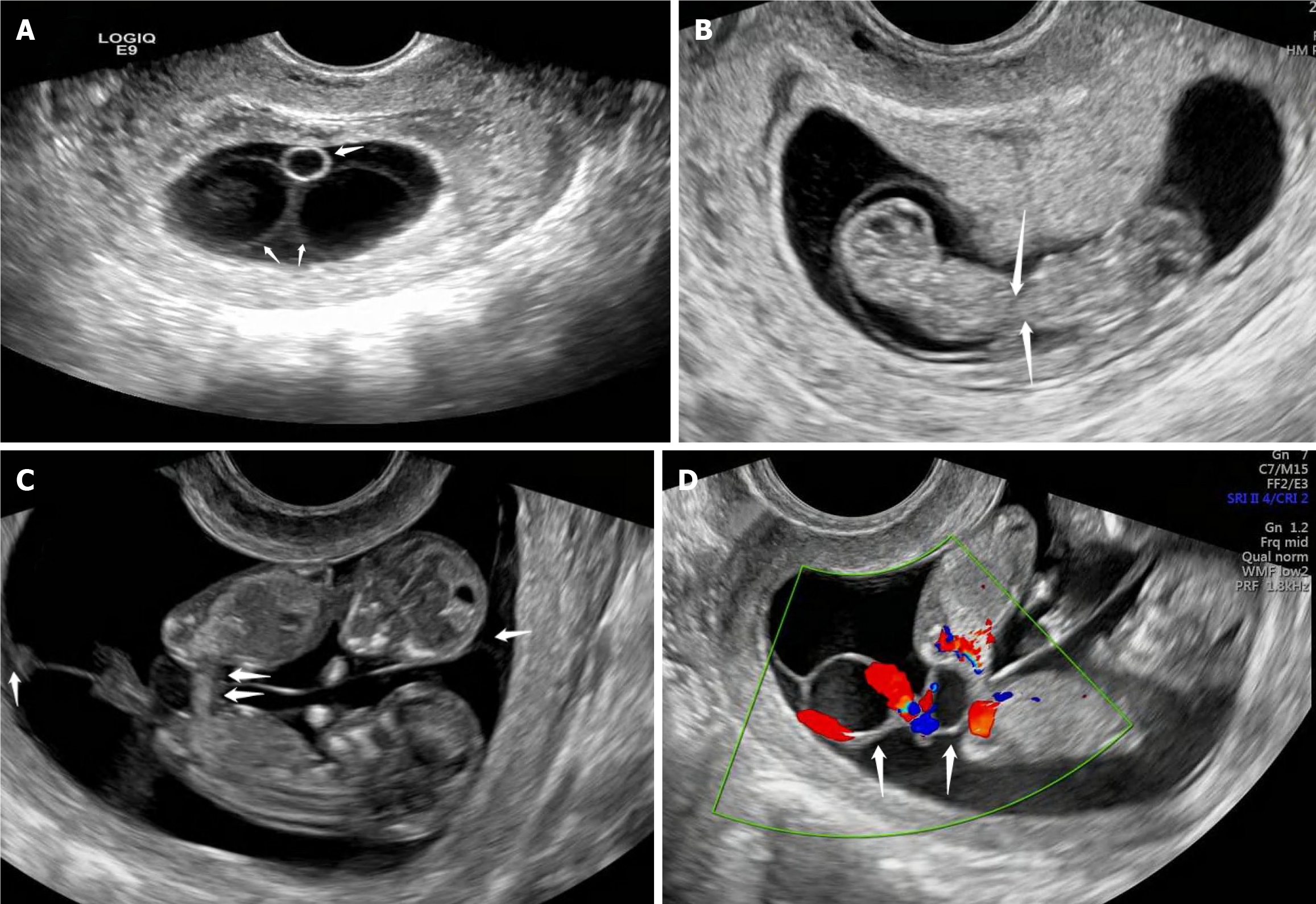
Figure 1 Transvaginal ultrasonography.
A: An amniotic septum is visible within the sac, and a yolk sac is visible at one end; B: An embryo and cardiac tube pulsation are observed in both amniotic sacs and the two embryos are connected in the lower abdomen under dynamic observation; C: Double fetuses with intestinal connection in the lower abdomen; D: Umbilical vessels intertwine to form a common umbilical cord at the site of twin attachment.
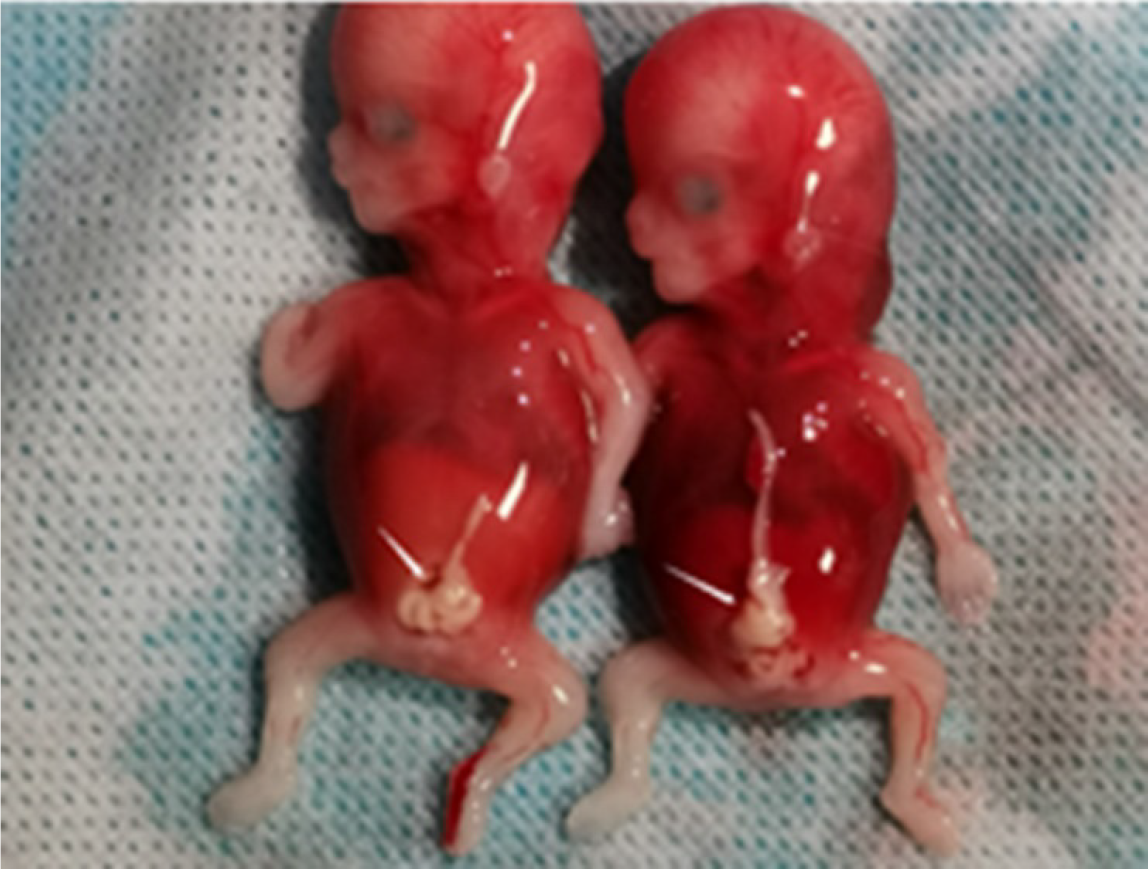
Figure 2 Poured postoperative.
Excretory tissues after induced abortion.
- Citation: Liang ZQ, Ding WQ. Twin fetuses associated with double amniotic sacs diagnosed using transvaginal ultrasonography: A case report. World J Clin Cases 2024; 12(18): 3534-3538
- URL: https://www.wjgnet.com/2307-8960/full/v12/i18/3534.htm
- DOI: https://dx.doi.org/10.12998/wjcc.v12.i18.3534


