Copyright
©The Author(s) 2024.
World J Clin Cases. Jun 26, 2024; 12(18): 3529-3533
Published online Jun 26, 2024. doi: 10.12998/wjcc.v12.i18.3529
Published online Jun 26, 2024. doi: 10.12998/wjcc.v12.i18.3529
Figure 1 Pathological section.
A: Microphotograph of laryngeal leiomyoma showing spindle cells arranged in fascicles and storiform pattern (Hematoxylin and eosin, × 10), microphotograph showing; B: Smooth-muscle actin; C: Desmin positivity.
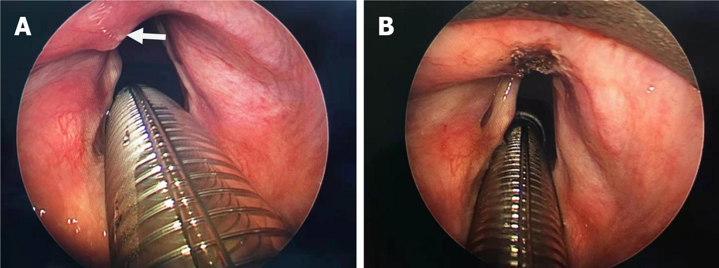
Figure 2 Laryngoscopy images.
A: Laryngoscopy imaging showing the solitary mass on the epiglottis; B: Intraoperatively, the mass located on the epiglottis was completely removed.
Figure 3 Timeline.
- Citation: Wu Y, Li JM, Zhang TJ, Wang X. Laryngeal leiomyoma: A case report and review of literature. World J Clin Cases 2024; 12(18): 3529-3533
- URL: https://www.wjgnet.com/2307-8960/full/v12/i18/3529.htm
- DOI: https://dx.doi.org/10.12998/wjcc.v12.i18.3529


