Copyright
©The Author(s) 2024.
World J Clin Cases. Jun 26, 2024; 12(18): 3351-3359
Published online Jun 26, 2024. doi: 10.12998/wjcc.v12.i18.3351
Published online Jun 26, 2024. doi: 10.12998/wjcc.v12.i18.3351
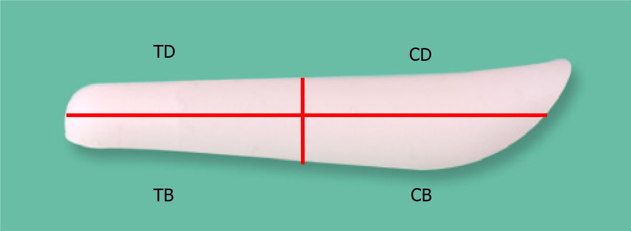
Figure 1 Four different compartments of the silicone nasal implant.
TD: Tip dorsum; TB: Tip base; CD: Cephalic dorsum; CB: Cephalic base.
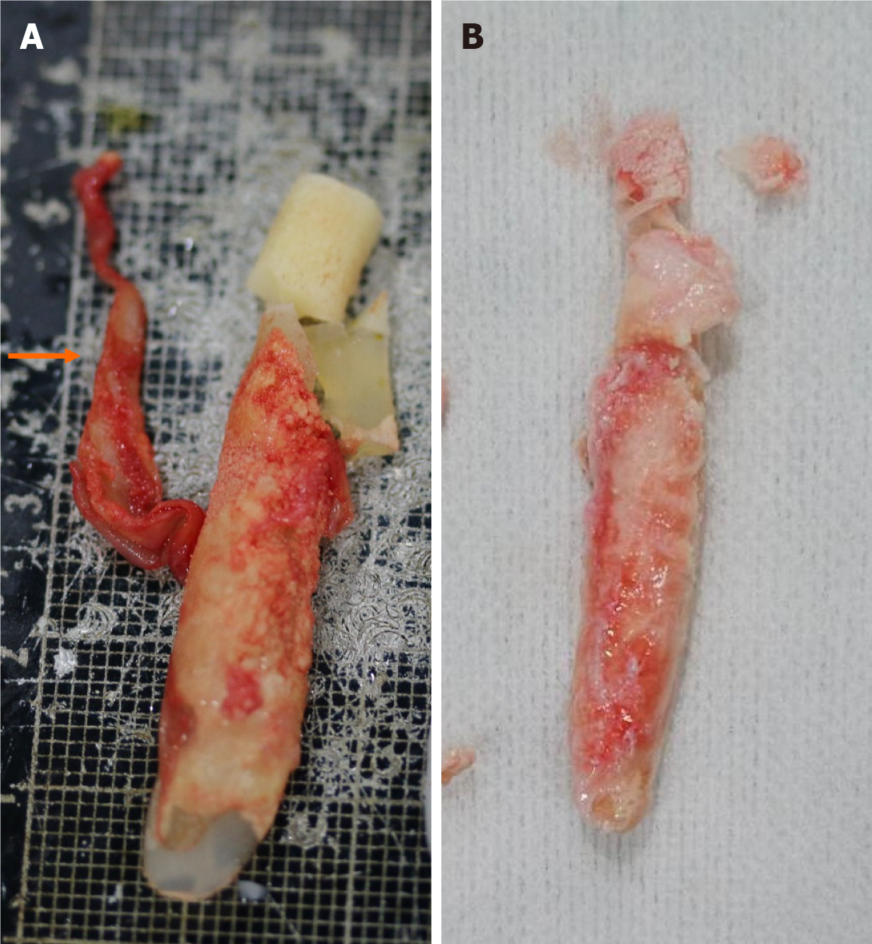
Figure 2 Gross findings of calcified nasal implant.
A: A calcified capsule (orange arrows) is observed around the implant; B: Another implant with severe multifocal peri-implant calcification.
Figure 3 Histopathologic findings of calcified nasal implant.
A: Calcified material surrounded by fibrotic tissue. Amorphous, irregular-shaped, basophilic deposits (orange arrowheads) suggesting dystrophic calcification were found surrounding the foreign material and fibrous tissue; B: Adjacent soft tissue exhibits a mild lymphocytic infiltration and a few foreign body-type giant cells (yellow arrowheads). Hematoxylin and Eosin staining, 500 μm.
Figure 4 Histopathologic findings of peri-implant tissue.
A: Dispersed multinucleated giant cells (orange arrowheads) in the surrounding fibrotic stroma; B: Sparse lymphocytic infiltration (yellow arrowheads) is also observed in the stroma. Hematoxylin and Eosin staining, 200 μm.
Figure 5 Computed tomography findings of calcified nasal implant.
A: A thick calcified capsule observed on the implant; B: Extensive peri-implant calcification surrounding with the right-side deviated implant.
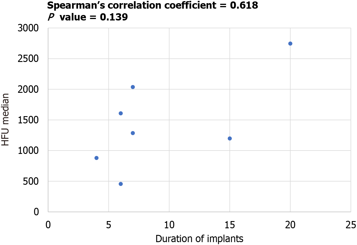
Figure 6 Relationship between the duration of the implant and the median value of Hounsfield unit.
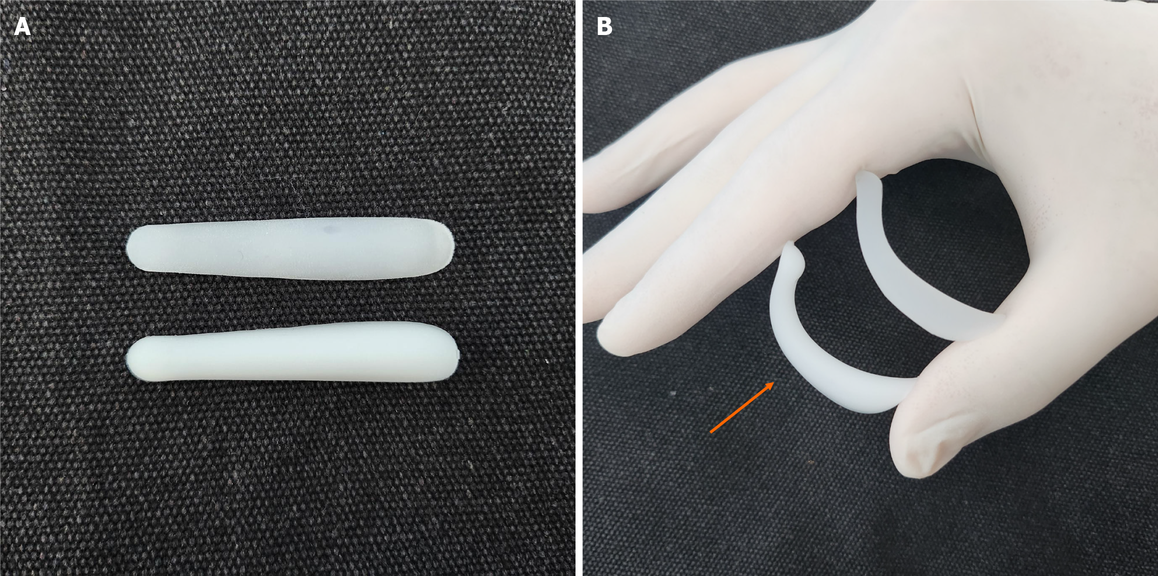
Figure 7 Bending strength test of soft silicone implant.
A: The implant on the above is a hard type implant (MISTI®; KEOSAN Trading, Seoul, Korea), and the implant on the below is a soft type nasal implant, (SOFTXIL®; BISTOOL, Seoul, Korea); B: It can be observed that the soft type nasal implant (left, orange arrow) bends much better when a mild grasping force is applied.
- Citation: Hwang YS, Kim TK, Yang DJ, Jang SH, Lee DW. Complicated calcified alloplastic implants in the nasal dorsum: A clinical analysis. World J Clin Cases 2024; 12(18): 3351-3359
- URL: https://www.wjgnet.com/2307-8960/full/v12/i18/3351.htm
- DOI: https://dx.doi.org/10.12998/wjcc.v12.i18.3351


