Copyright
©The Author(s) 2024.
World J Clin Cases. Jun 26, 2024; 12(18): 3304-3313
Published online Jun 26, 2024. doi: 10.12998/wjcc.v12.i18.3304
Published online Jun 26, 2024. doi: 10.12998/wjcc.v12.i18.3304
Figure 1 Endoscopic view of the colon.
A: The picture shows a complete loss of haustration and the presence of yellow matter and blood (as an expression of necrosis); B: Complete loss of haustration, absence of vascular pattern, and mucosal glare are also seen; C: Endoscopic view of the ascending colon. The presence of a tumor mass in the ascending colon completely disrupts the structure of the large intestine. Necrosis is clearly visible in the very center, while partial haustration can still be observed in the periphery.
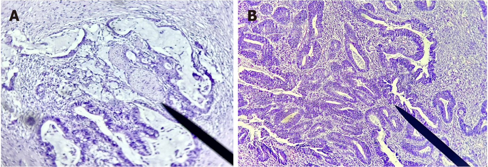
Figure 2 Mucinous adenocarcinoma of the colon, with perineural invasion (marked by an arrow).
A: The World Health Organization recognized a subtype of colorectal carcinoma with mucin lakes comprising at least 50% of a tumor mass (HE staining, × 100); B: Invasive moderately differentiated adenocarcinoma of the colon (HE staining, × 100).
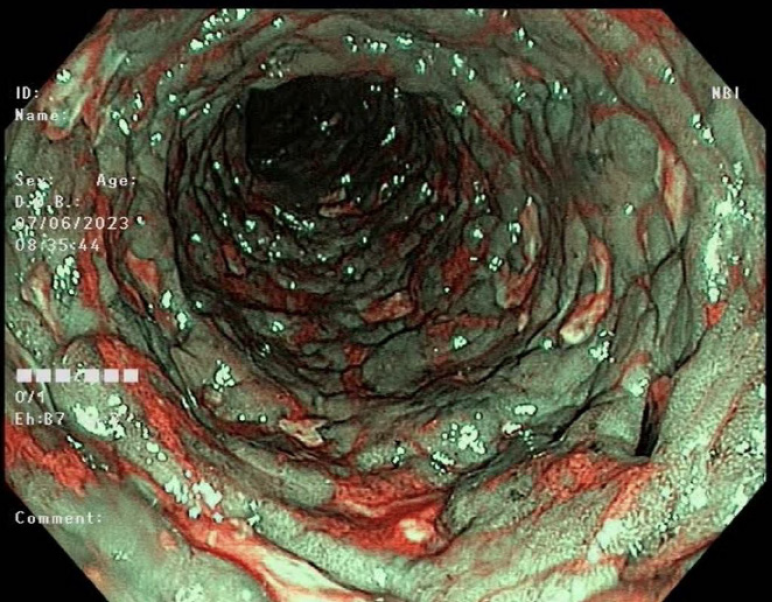
Figure 3 Image showing a mucous membrane with pronounced pavement (a typical picture of Crohn's disease).
Narrow band imaging allows looking for visible signs of malignancy - a disturbed pattern, lack of regularity, irregular shape, dark spots in the crypts, and unclear borders. Mucus and residual intestinal contents are represented in red (poor preparation). Edematous mucosa is in blue with a regular pit pattern. Macroscopic signs of malignancy are absent.
- Citation: Gulinac M, Kiprin G, Tsranchev I, Graklanov V, Chervenkov L, Velikova T. Clinical issues and challenges in imaging of gastrointestinal diseases: A minireview and our experience. World J Clin Cases 2024; 12(18): 3304-3313
- URL: https://www.wjgnet.com/2307-8960/full/v12/i18/3304.htm
- DOI: https://dx.doi.org/10.12998/wjcc.v12.i18.3304


