Copyright
©The Author(s) 2024.
World J Clin Cases. Jun 16, 2024; 12(17): 3265-3270
Published online Jun 16, 2024. doi: 10.12998/wjcc.v12.i17.3265
Published online Jun 16, 2024. doi: 10.12998/wjcc.v12.i17.3265
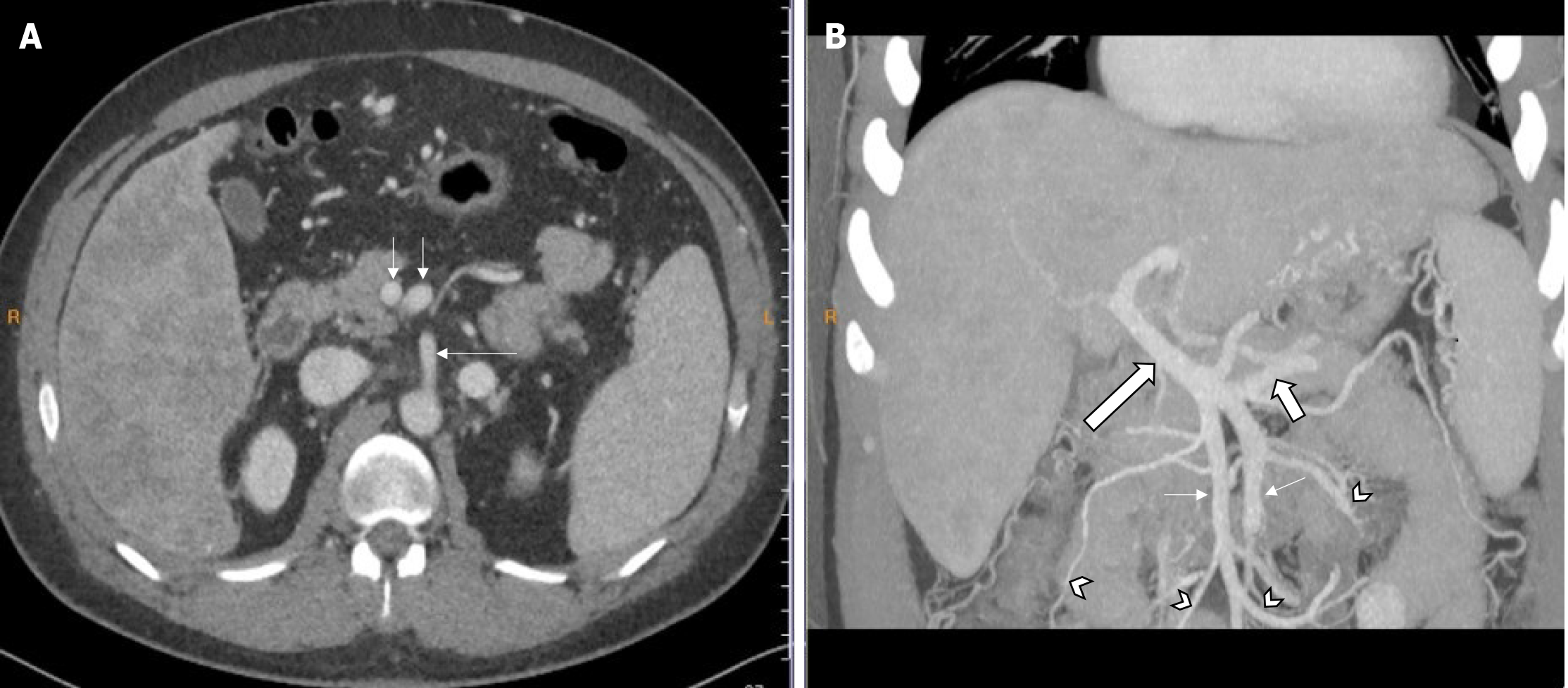
Figure 1 An enhanced multidetector computed tomography of the upper abdomen and abdominal angiography images from a 34-year-old male patient with cirrhosis.
A: A cross-sectional venous phase enhanced image showed parallel double superior mesenteric veins (SMVs) (short arrow) in front of the superior mesenteric artery (SMA) (long arrow), the left SMV is located in front of the SMA, and the right SMV is located on the right front side of the SMA; B: A maximum intensity projection reconstruction image of abdominal angiography showed the left and right SMV (thin arrow) join the portal vein (long thick arrow) in parallel together with the splenic vein (short thick arrow). The branches of the left SMV mainly receive the blood from the proximal small intestine, and the branches of the right SMV mainly receive the blood from the distal small intestine and the proximal colon (V-shaped arrow).
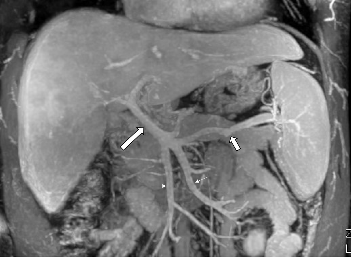
Figure 2 An abdominal magnetic resonance venography of a 34-year-old male patient with cirrhosis.
The magnetic resonance venography images showed the left and right superior mesenteric vein (thin arrow) join the portal vein (long thick arrow) in parallel together with the splenic vein (short thick arrow).
- Citation: Tang W, Peng S. Multidetector computer tomography and magnetic resonance imaging of double superior mesenteric veins: A case report. World J Clin Cases 2024; 12(17): 3265-3270
- URL: https://www.wjgnet.com/2307-8960/full/v12/i17/3265.htm
- DOI: https://dx.doi.org/10.12998/wjcc.v12.i17.3265


