Copyright
©The Author(s) 2024.
World J Clin Cases. Jun 16, 2024; 12(17): 3235-3242
Published online Jun 16, 2024. doi: 10.12998/wjcc.v12.i17.3235
Published online Jun 16, 2024. doi: 10.12998/wjcc.v12.i17.3235
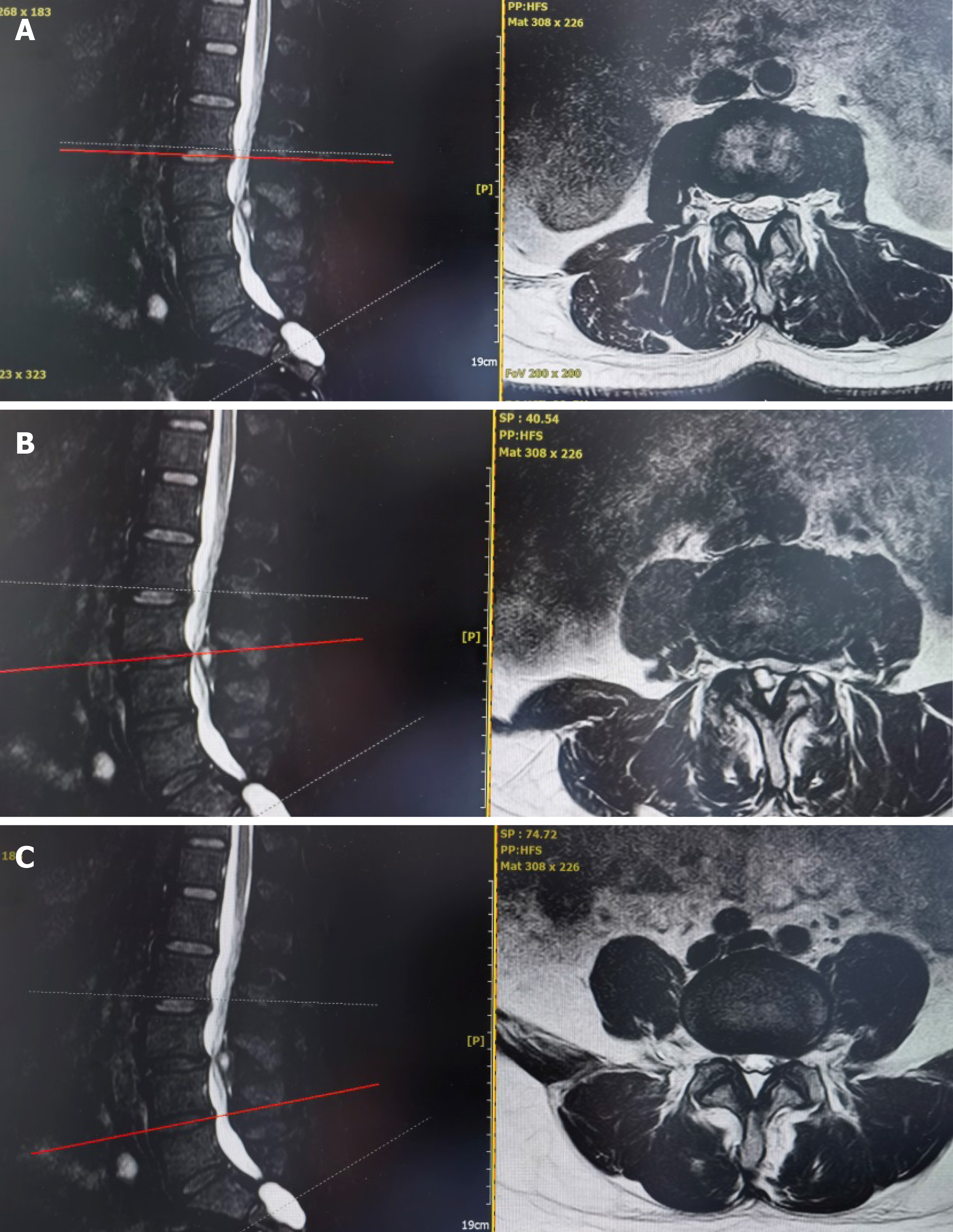
Figure 1 Preoperative data of patients.
A: L2-3 intervertebral disc protruded to the right rear, L3 vertebral body slipped forward I°; B: L3-4 intervertebral discs were deformed and bulged; C: L4-5 intervertebral discs were deformed and bulged.
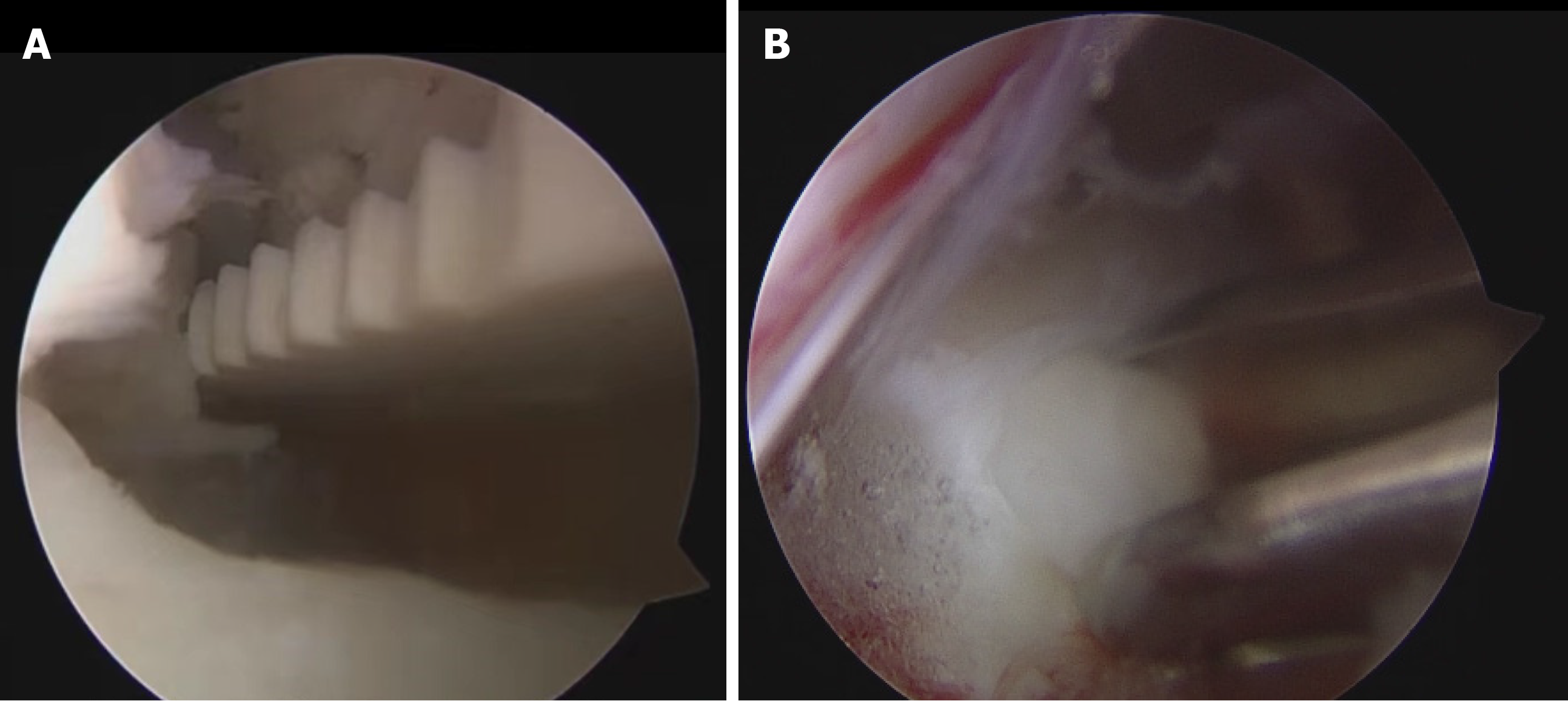
Figure 2 Unilateral biportal endoscopy.
A: Patient surgical data - intraoperative unilateral biportal endoscopy nerve decompression; B: Unilateral biportal endoscopy microscopic fusion.
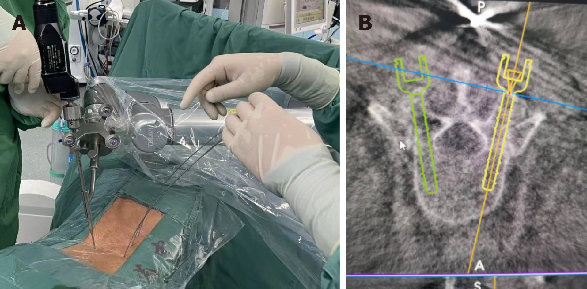
Figure 3 Robot assisted pedicle screw implantation.
A: Robot operation; B: Good screw position.
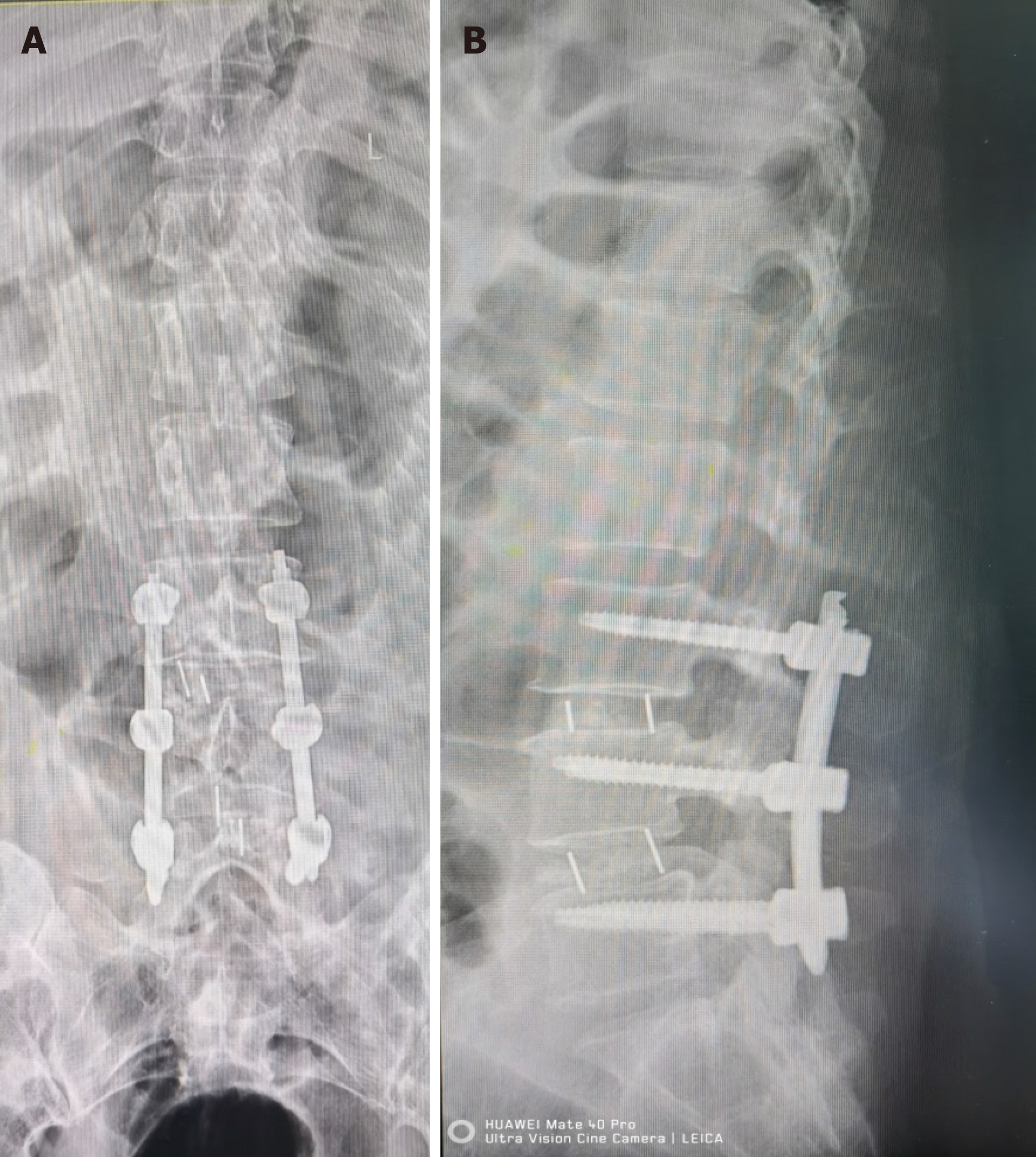
Figure 4 Postoperative data.
A: The lumbar spine radiograph showed that the screw was in good position; B: Lumbar lateral film shows good screw position.
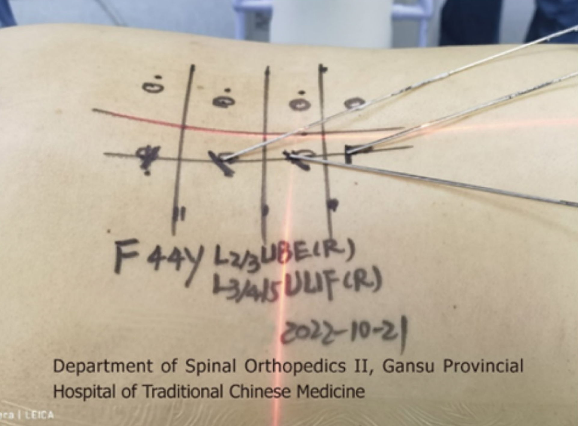
Figure 5
Patient surgical data - preoperative body surface positioning.
- Citation: Liu YD, Xu DF, Deng Q, Zhang YJ, Guo TF, Peng RD, Li JJ. Treatment of lumbar disc herniation with robot combined with unilateral biportal endoscopic technology: A case report. World J Clin Cases 2024; 12(17): 3235-3242
- URL: https://www.wjgnet.com/2307-8960/full/v12/i17/3235.htm
- DOI: https://dx.doi.org/10.12998/wjcc.v12.i17.3235


