Copyright
©The Author(s) 2024.
World J Clin Cases. Jun 16, 2024; 12(17): 3226-3234
Published online Jun 16, 2024. doi: 10.12998/wjcc.v12.i17.3226
Published online Jun 16, 2024. doi: 10.12998/wjcc.v12.i17.3226
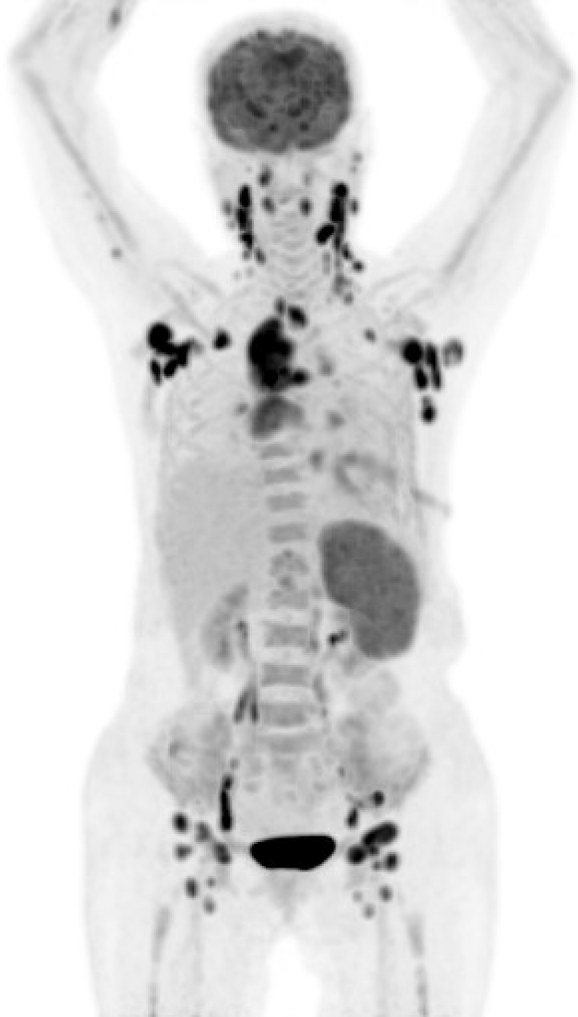
Figure 1 Positron-emission tomography-computed tomography scan.
Initial positron-emission tomography-computed tomography showed malignant lymphoma activity disseminated in the Waldeyer's ring, bilateral nuchal, bilateral submental and submandibular, bilateral neck, bilateral supraclavicular, bilateral mediastinal, bilateral pulmonary hilar, bilateral axillary, splenic, celiac, paraaortic, bilateral iliac, and bilateral inguinal lymph nodes.
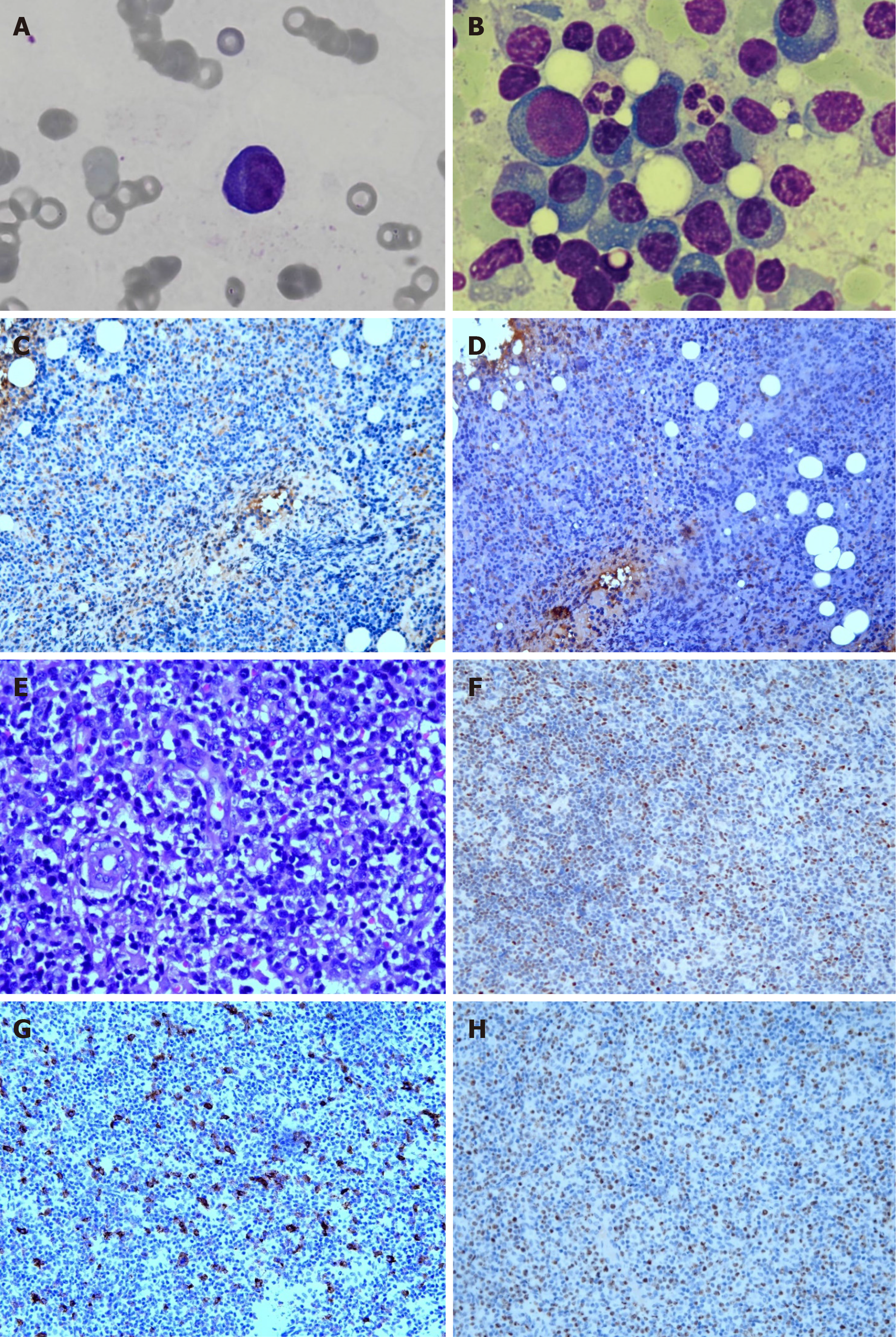
Figure 2 Microscopic evaluation of the lesion.
A: A plasma cell and rouleaux formation in peripheral blood smear (magnification, 1000 ×); B: Plasmacytosis with atypical and immature plasma cells in bone marrow smear (magnification, 1000 ×); C: Immunohistochemistry (IHC) results for kappa show positivity in bone marrow (magnification, 200 ×); D: IHC results for lambda show positivity in bone marrow (magnification, 200 ×); E: Small- to medium-sized atypical lymphocytes and follicular dendritic cells with clear cytoplasm and capillaries with enlarged endothelial cells are observed using hematoxylin and eosin staining (magnification, 400 ×); F: IHC results for BCL-6 show positivity (magnification, 100 ×); G: IHC results for CD30 show positivity (magnification, 100 ×); H: IHC results for Ki-67 show positivity (magnification, 100 ×).
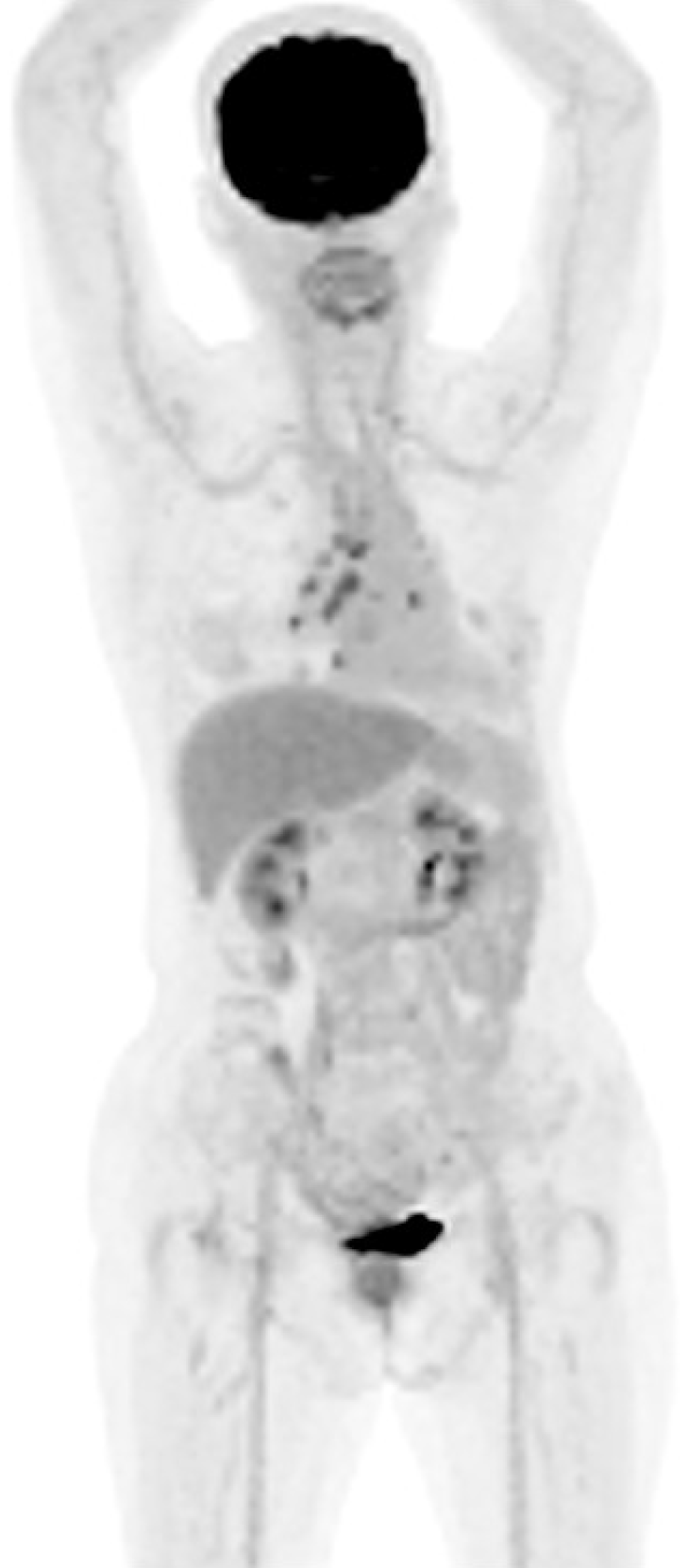
Figure 3 Positron-emission tomography-computed tomography scan.
Follow-up positron-emission tomography-computed tomography revealed residual lymphoma in the mediastinum and bilateral pulmonary hila.
- Citation: Lin CC, Lee HL, Chuo HY, Chen TA, Liu MY, Chen LM. Plasmacytosis mimicking multiple myeloma in angioimmunoblastic T-cell lymphoma: A case report and review of literature. World J Clin Cases 2024; 12(17): 3226-3234
- URL: https://www.wjgnet.com/2307-8960/full/v12/i17/3226.htm
- DOI: https://dx.doi.org/10.12998/wjcc.v12.i17.3226


