Copyright
©The Author(s) 2024.
World J Clin Cases. Jun 16, 2024; 12(17): 3194-3199
Published online Jun 16, 2024. doi: 10.12998/wjcc.v12.i17.3194
Published online Jun 16, 2024. doi: 10.12998/wjcc.v12.i17.3194
Figure 1 Ultrasonography image of the pseudoaneurysm (arrowheads) in the left superficial brachial artery.
A: Short axis view; B: Long axis view.
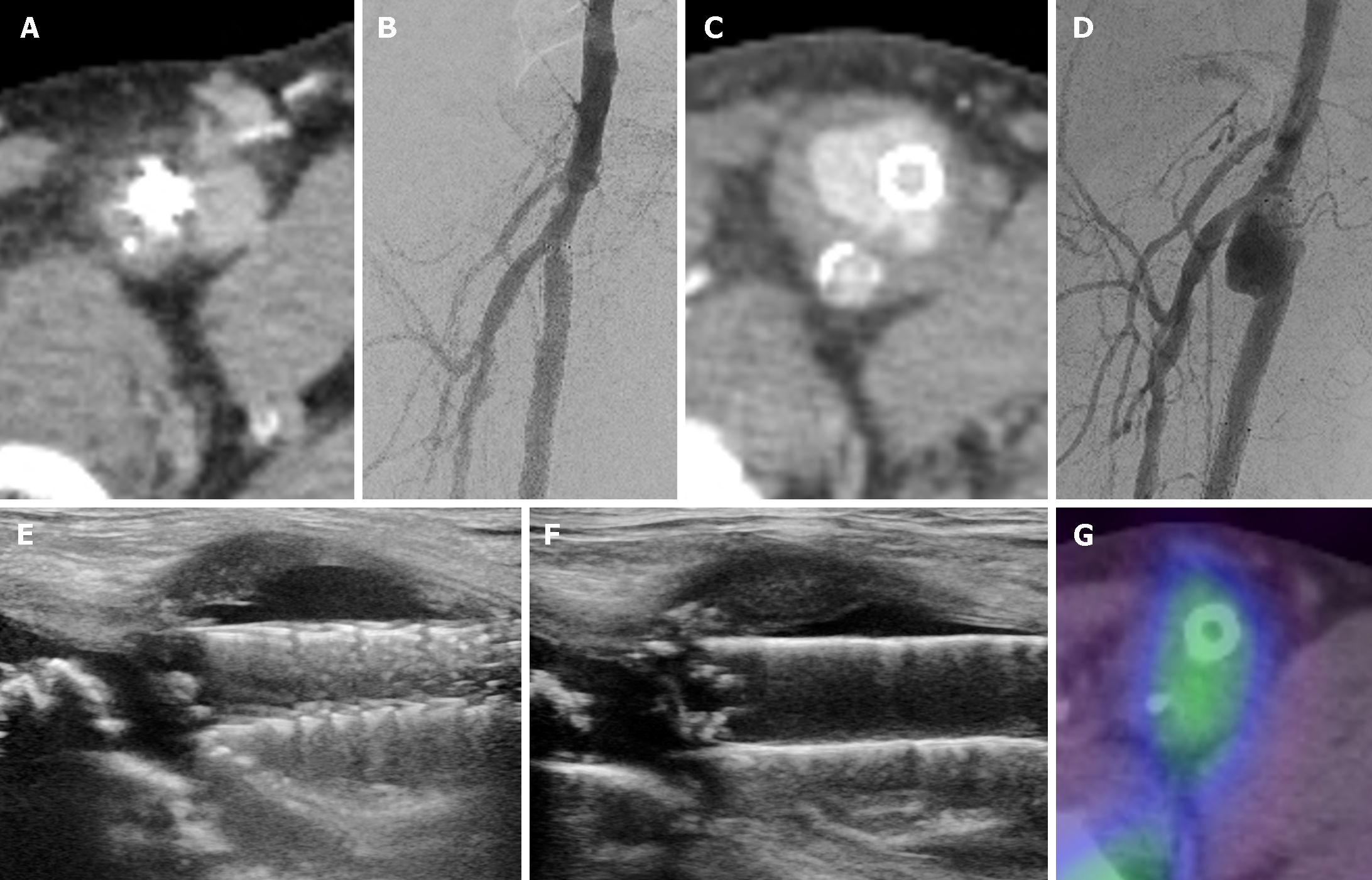
Figure 2 Pseudoaneurysm in the right superficial femoral artery.
A: Contrast-enhanced computed tomography (CT) image at admission; B: Angiogram image at admission; C: Contrast-enhanced CT image 7 wk after admission; D: Angiogram image 7 wk after admission showing obvious enlargement within a short period of time; E: Ultrasonographic images at 3 months; F: Ultrasonographic images at 6 months from the date of admission. Although there is no significant change, there is a slight increasing trend; G: Gallium scintigraphy 3 wk after admission reveals slight uptake.
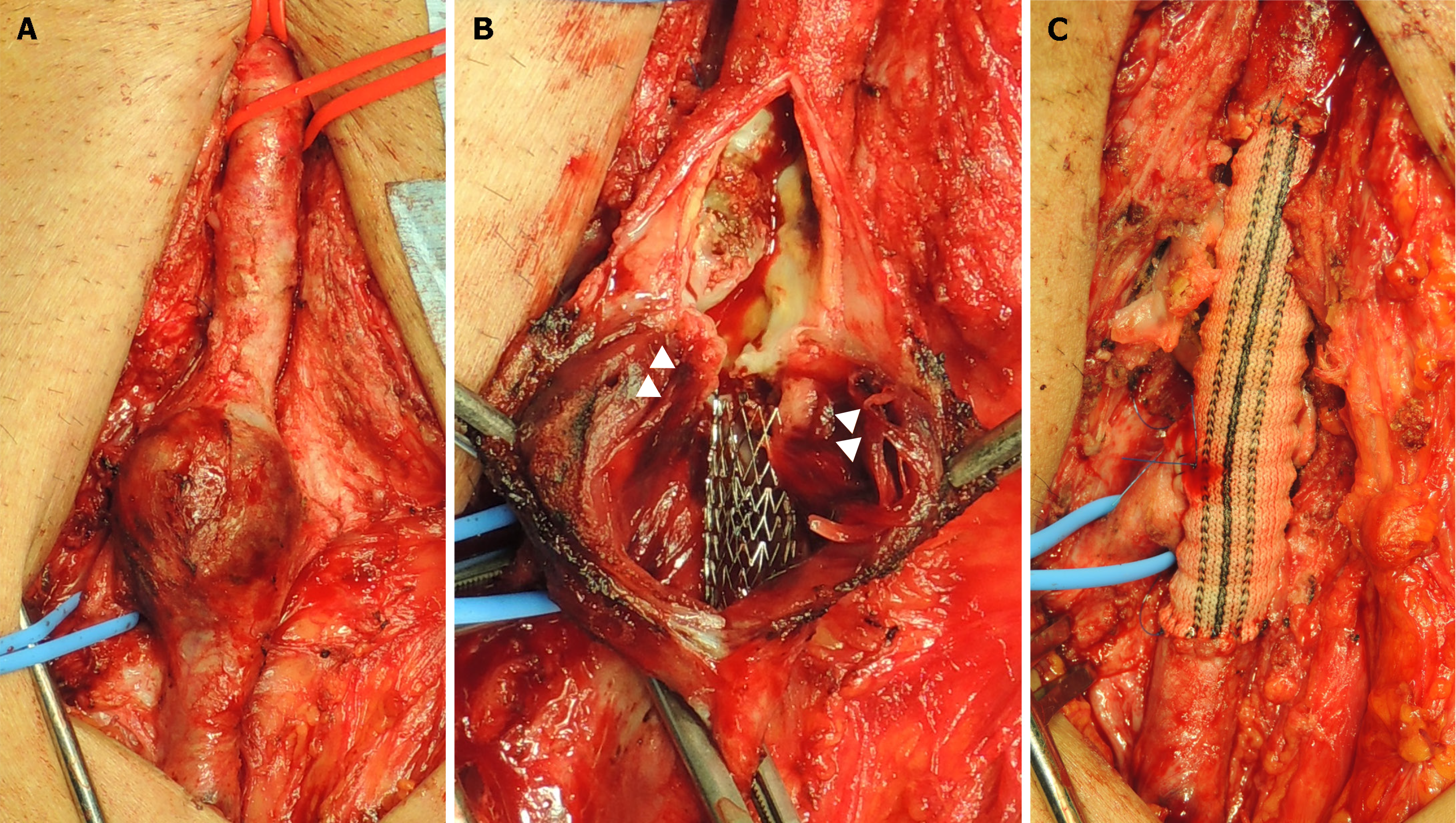
Figure 3 Intraoperative photographic images.
A: Common femoral artery, superficial femoral artery, and deep femoral artery are isolated. The aneurysm is visible at the bifurcation of the femoral arteries; B: Each artery is clamped and the aneurysm was incised. The stent can be observed inside the blood vessel wall (arrowheads); C: Vascular reconstruction is performed using a rifampicin-soaked Dacron graft.
- Citation: Akai T, Ninomiya S, Kaneko T. Superficial femoral artery pseudoaneurysm at implantation site of drug eluting stent discovered due to bacteremia: A case report. World J Clin Cases 2024; 12(17): 3194-3199
- URL: https://www.wjgnet.com/2307-8960/full/v12/i17/3194.htm
- DOI: https://dx.doi.org/10.12998/wjcc.v12.i17.3194


