Copyright
©The Author(s) 2024.
World J Clin Cases. Jun 16, 2024; 12(17): 3144-3150
Published online Jun 16, 2024. doi: 10.12998/wjcc.v12.i17.3144
Published online Jun 16, 2024. doi: 10.12998/wjcc.v12.i17.3144
Figure 1 Olecranon fracture and evolution of myositis ossificans after internal fixation.
A: 3D reconstruction shows comminuted fracture of the scaphoid bone; B: 3D after surgery; C: 6 wk after surgery, ossifying myositis appeared; D: 10 wk after surgery, evidence of myositis ossificans.
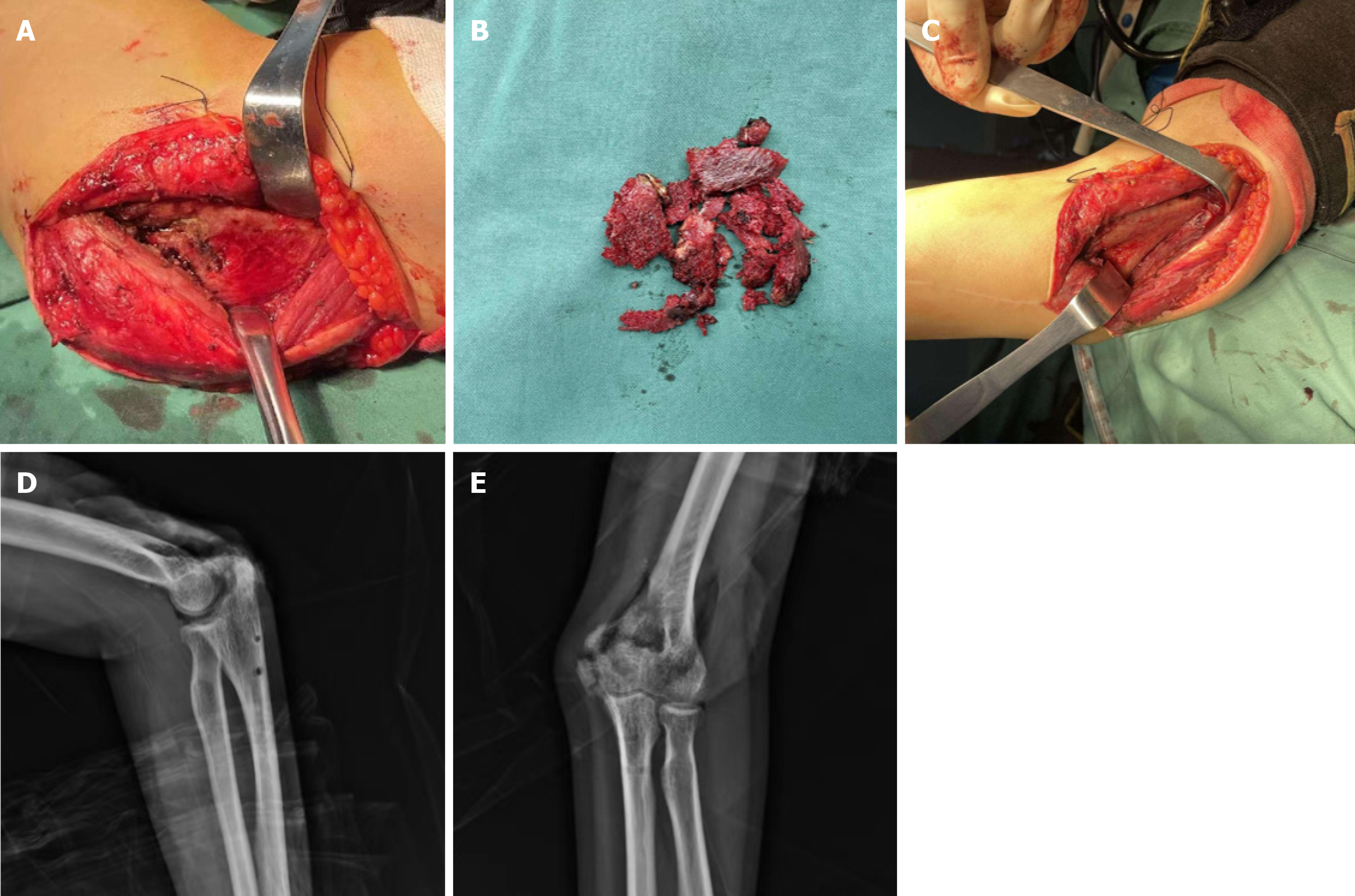
Figure 2 Clearance of myositis ossificans during surgery and measurement of joint motion.
A: The surgical field was adequately exposed before removing the bone spur; B: The bone spur was removed during surgery; C: Visual field of elbow joint after removal of bone spurs; D: Measurement of elbow joint range of motion: Elbow flexion was 150; E: Elbow extension was 20.
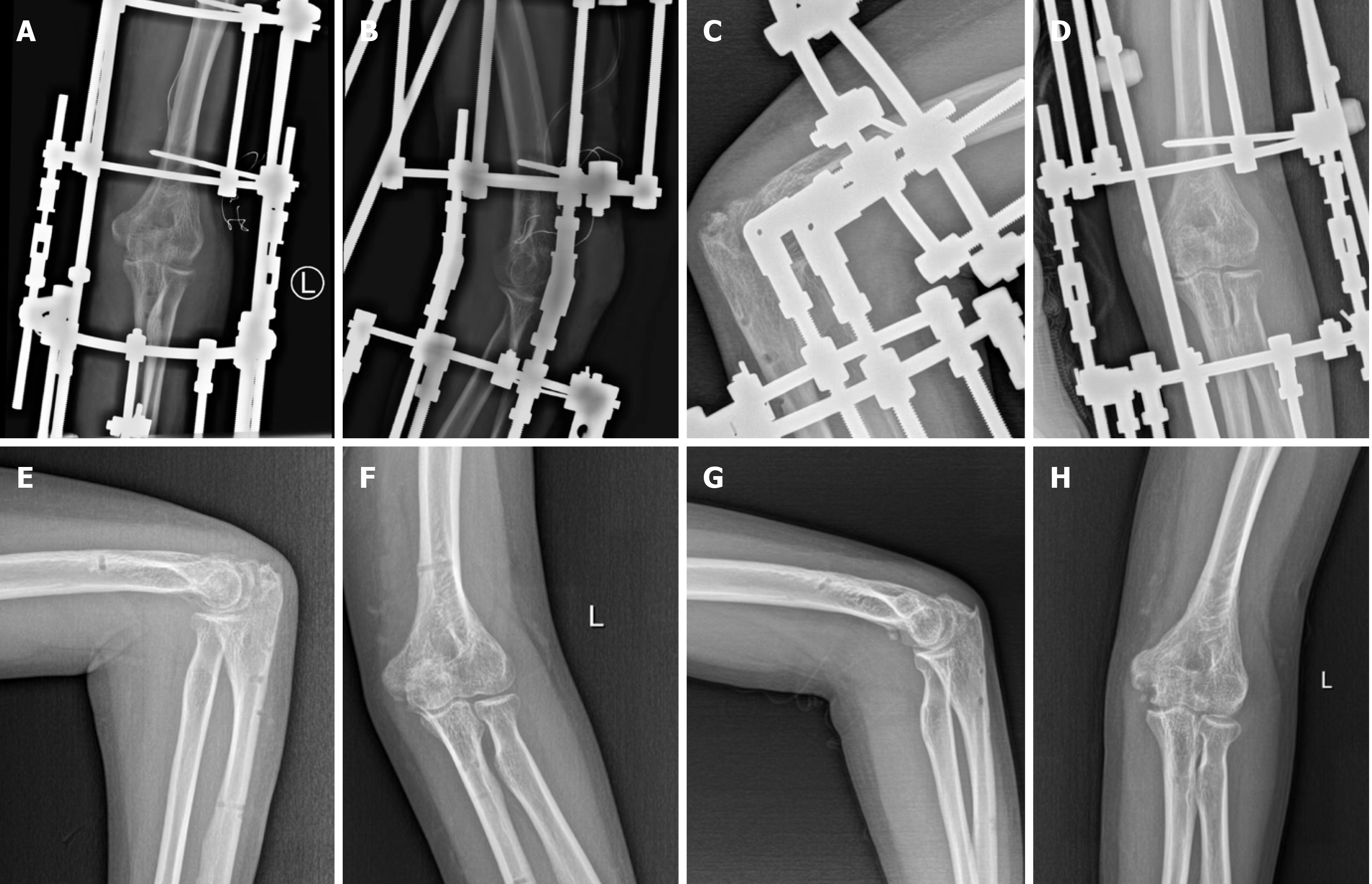
Figure 3 After the Ilizarov frame was installed and removed.
A and B: 2 d postoperatively; C and D: 6 wk postoperatively; E and F: 8 wk postoperatively; G and H: 8 months postoperatively.
- Citation: Zhou MW, Zhang PW, Zhang AL, Wei CH, Xu YD, Chen W, Fu ZB. Ilizarov technique for treating elbow stiffness caused by myositis ossificans: A case report. World J Clin Cases 2024; 12(17): 3144-3150
- URL: https://www.wjgnet.com/2307-8960/full/v12/i17/3144.htm
- DOI: https://dx.doi.org/10.12998/wjcc.v12.i17.3144


