Copyright
©The Author(s) 2024.
World J Clin Cases. Jun 16, 2024; 12(17): 3123-3129
Published online Jun 16, 2024. doi: 10.12998/wjcc.v12.i17.3123
Published online Jun 16, 2024. doi: 10.12998/wjcc.v12.i17.3123
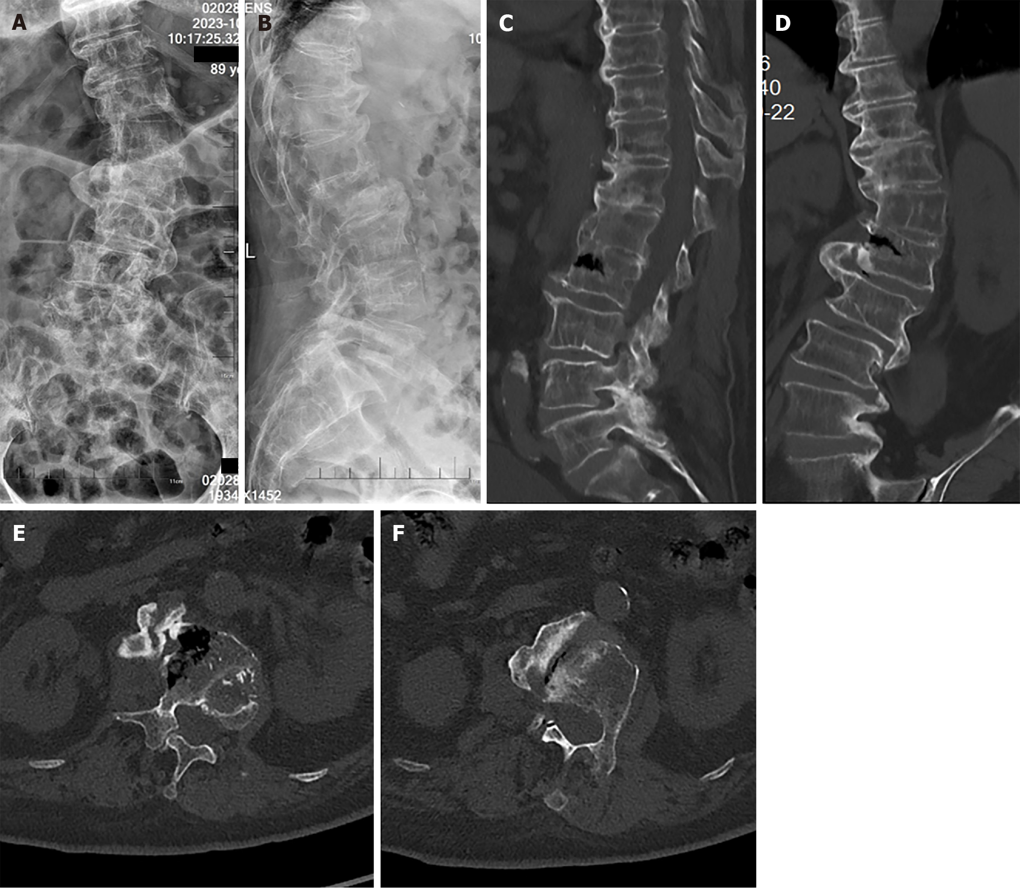
Figure 1 Preoperative imaging examination of patient.
A: Anteroposterior X-ray of lumbar vertebrae; B: Lateral X-ray of lumbar vertebrae; C: Computed tomography (CT) sagittal reconstruction of lumbar vertebrae; D: CT coronal reconstruction of lumbar vertebrae; E and F: CT cross section scan of L2 fractured vertebral body.
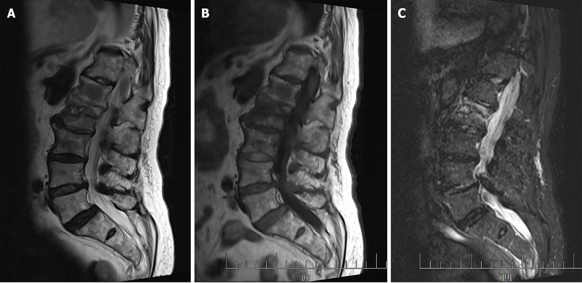
Figure 2 Preoperative magnetic resonance imaging examination of the patient.
A: T2-weighted imaging of lumbar vertebrae; B: T1-weighted imaging of lumbar vertebrae; C: Short tau inversion recovery sequence of lumbar vertebrae.
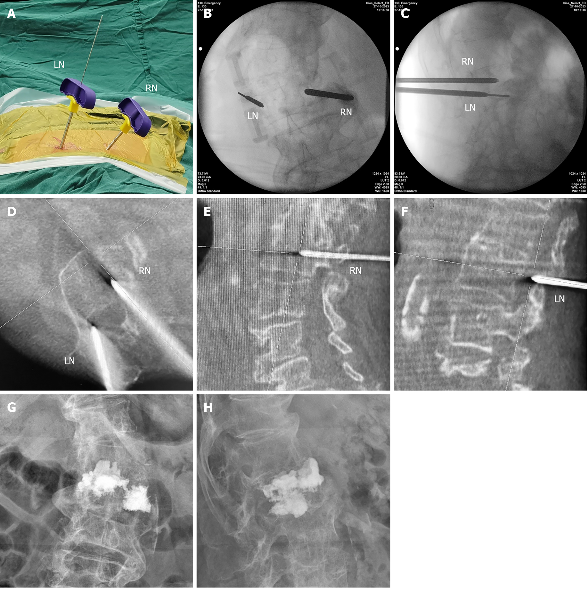
Figure 3 Surgical procedure.
A: Intraoperative photograph; B: Anteroposterior X-ray of positioning needles; C: Lateral X-ray of positioning needles; D: Intraoperative O-arm scan of intravertebral position of positioning needles; E: Position of the right positioning needle in the vertebra in sagittal reconstruction; F: Position of the left positioning needle in the vertebra in sagittal reconstruction; G: Postoperative anteroposterior X-ray of lumbar vertebrae; H: Postoperative lateral X-ray of lumbar vertebrae. LN: Left needle; RN: Right needle.
- Citation: Saijilafu, Zhou JW, Wang GL, Sun KH, Xie JL. Percutaneous kyphoplasty in the treatment of Kümmell disease in lumbar scoliosis: A case report. World J Clin Cases 2024; 12(17): 3123-3129
- URL: https://www.wjgnet.com/2307-8960/full/v12/i17/3123.htm
- DOI: https://dx.doi.org/10.12998/wjcc.v12.i17.3123


