Copyright
©The Author(s) 2024.
World J Clin Cases. Jun 6, 2024; 12(16): 2904-2910
Published online Jun 6, 2024. doi: 10.12998/wjcc.v12.i16.2904
Published online Jun 6, 2024. doi: 10.12998/wjcc.v12.i16.2904
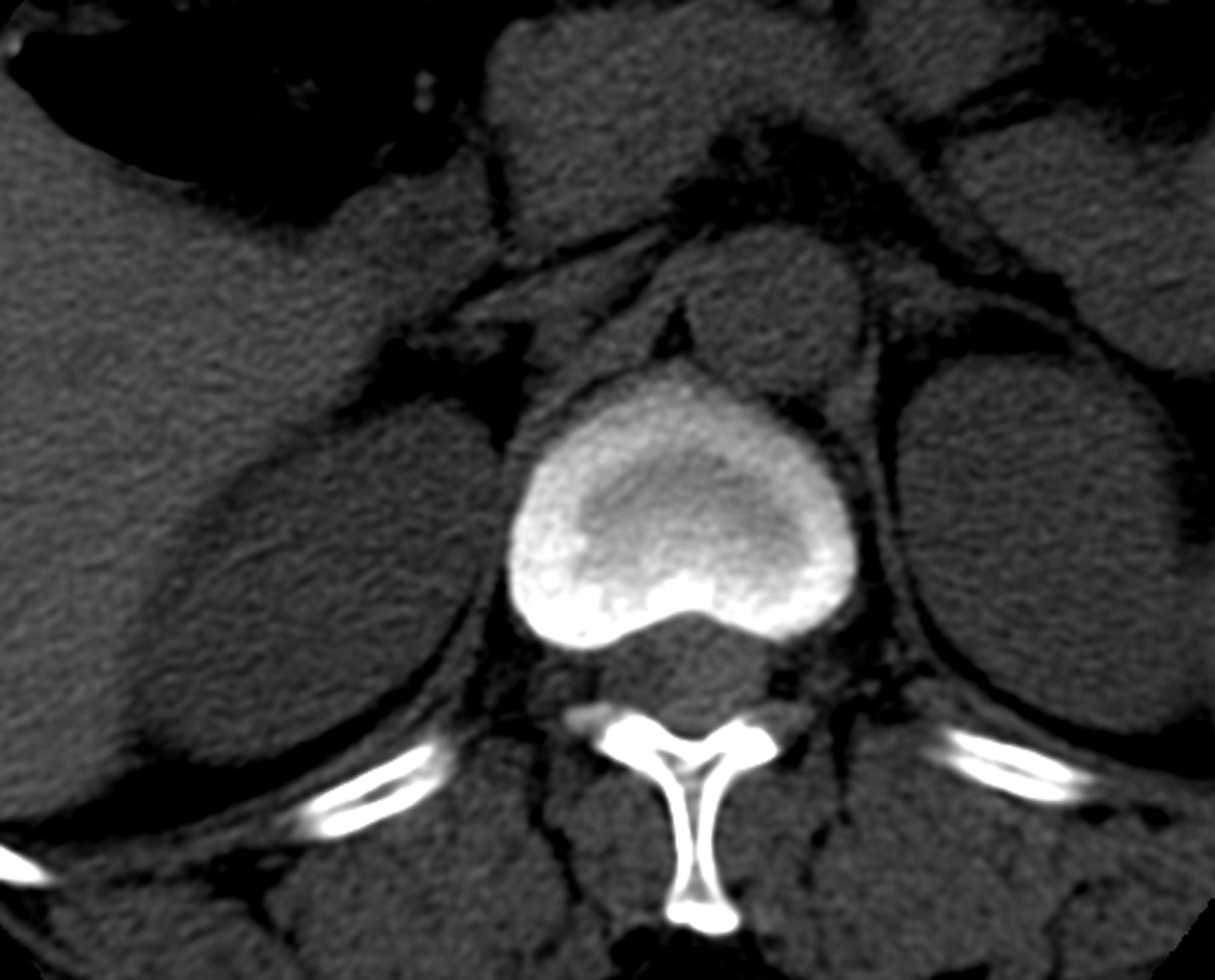
Figure 1 Computed tomography showing that the spinal canal at thoracic 12 to lumbar 1 had a slightly greater nodular density and an unclear boundary.
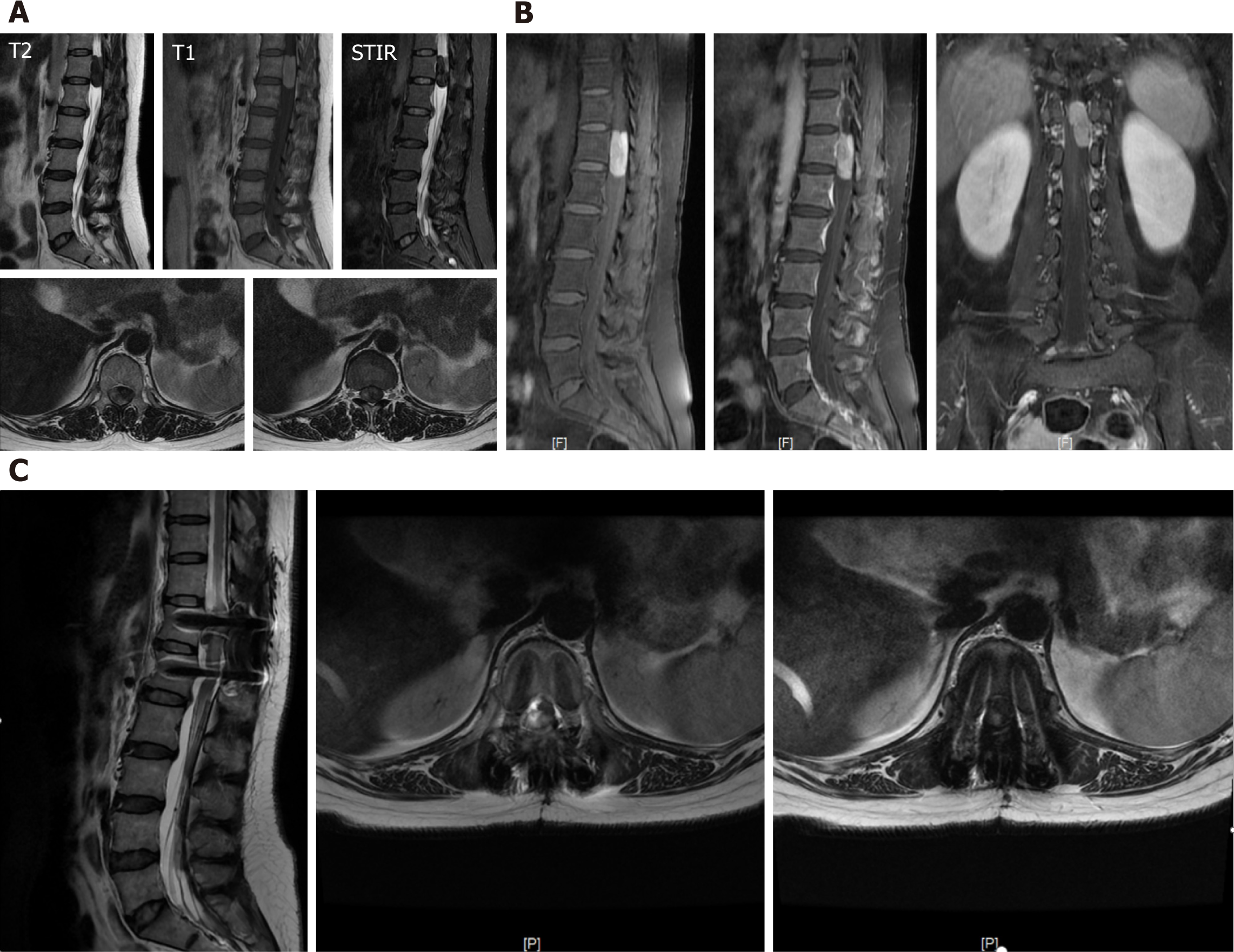
Figure 2 Magnetic resonance imaging and enhanced magnetic resonance image.
A: Magnetic resonance imaging revealed spindle-shaped swelling in the spinal canal of thoracic 12 to lumbar 1, with a slightly greater signal in T1-weighted imaging and an equal signal in T1-weighted imaging with a clear boundary; B: Enhanced magnetic resonance image showing that the number of foci was significantly greater; C: An abnormal signal was still observed in the spinal canal after surgery, indicating residual tumor tissue.
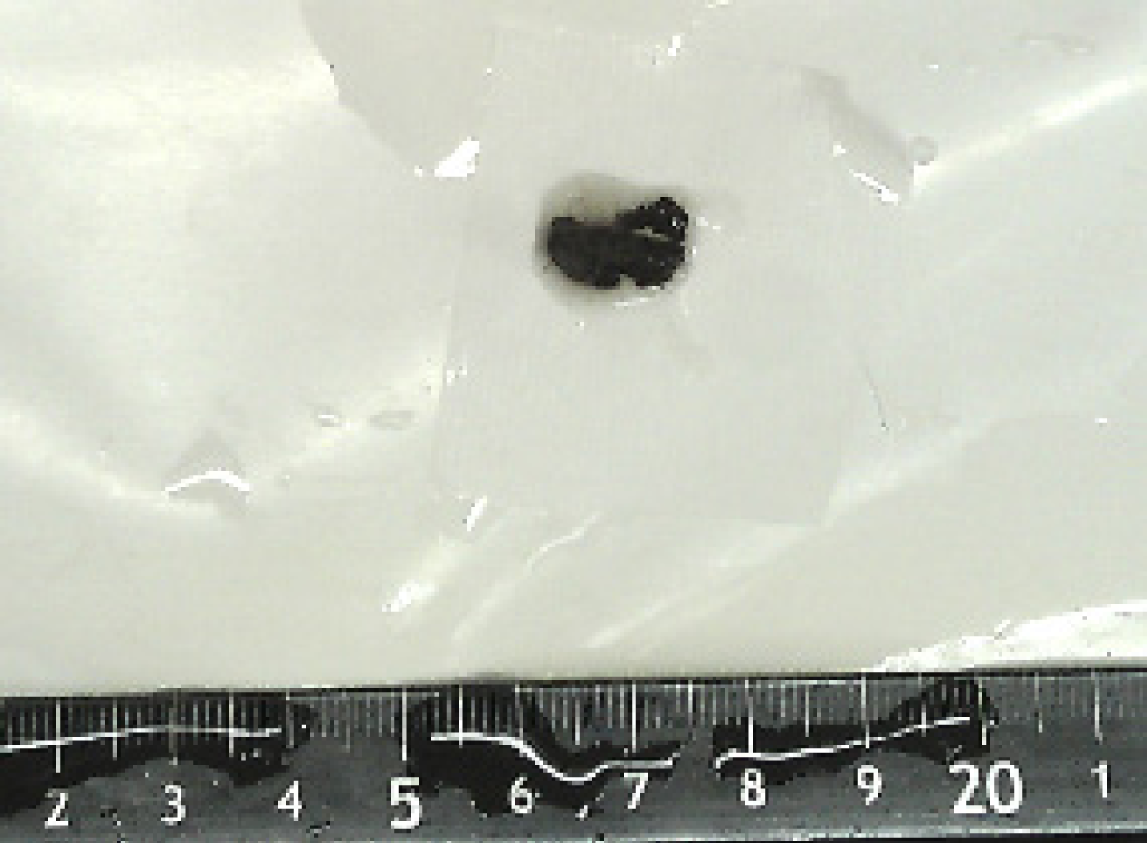
Figure 3 The mass is generally black-brown and clot-shaped.
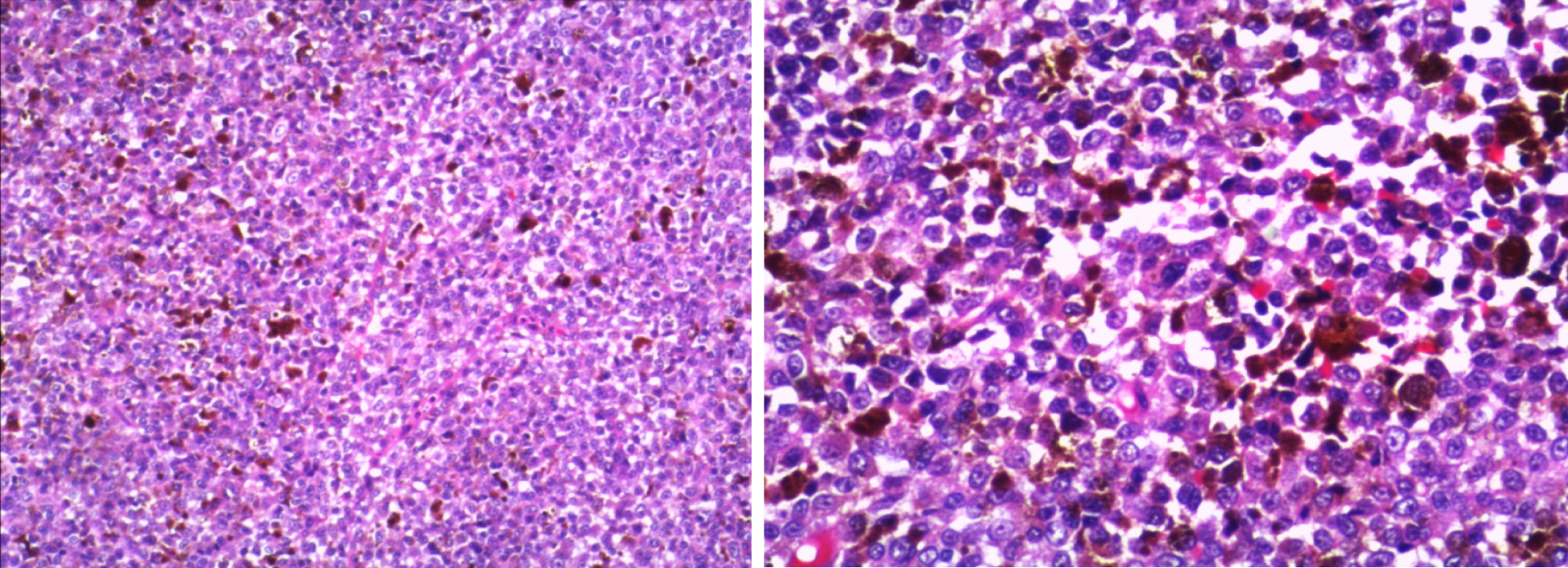
Figure 4 The tumor cells have a nest sheet and are rich in pigment, with tumor cell atypia, large nuclei, obvious nucleoli, and visible nuclear division, consistent with malignant melanoma.
- Citation: Huang JB, Xue HJ, Zhu BY, Lei Y, Pan L. Primary thoracolumbar intraspinal malignant melanoma: A case report. World J Clin Cases 2024; 12(16): 2904-2910
- URL: https://www.wjgnet.com/2307-8960/full/v12/i16/2904.htm
- DOI: https://dx.doi.org/10.12998/wjcc.v12.i16.2904


