Copyright
©The Author(s) 2024.
World J Clin Cases. Jun 6, 2024; 12(16): 2869-2875
Published online Jun 6, 2024. doi: 10.12998/wjcc.v12.i16.2869
Published online Jun 6, 2024. doi: 10.12998/wjcc.v12.i16.2869
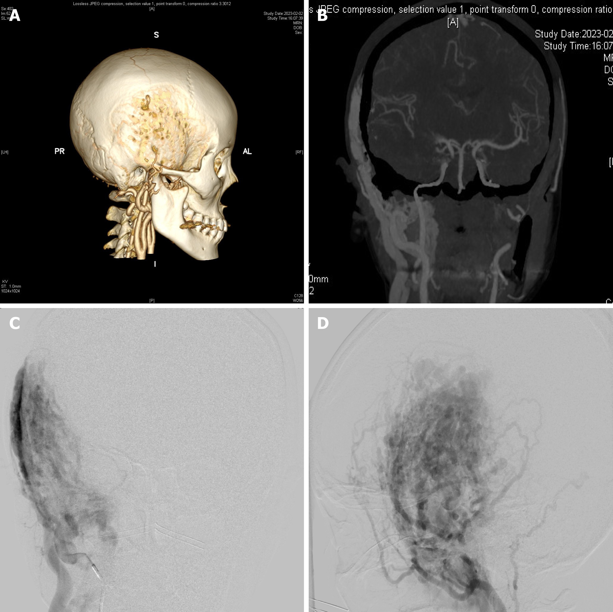
Figure 1 Preoperative cerebral angiography and computed tomography angiography of cerebral arteries.
A: The right temporal bone was extensively invaded and became worm-like changes; B-D: Cerebral angiography and computed tomography angiography of the cerebral artery show multiple tortuous vascular shadows in the right temporal subcutis, with blood supply from the right external carotid artery and lesions invading the adjacent cranial eminence to the epidural. External carotid artery angiography showed a hemangioma supplied by maxillary and superficial temporal arteries, size about 10 cm × 6 cm.
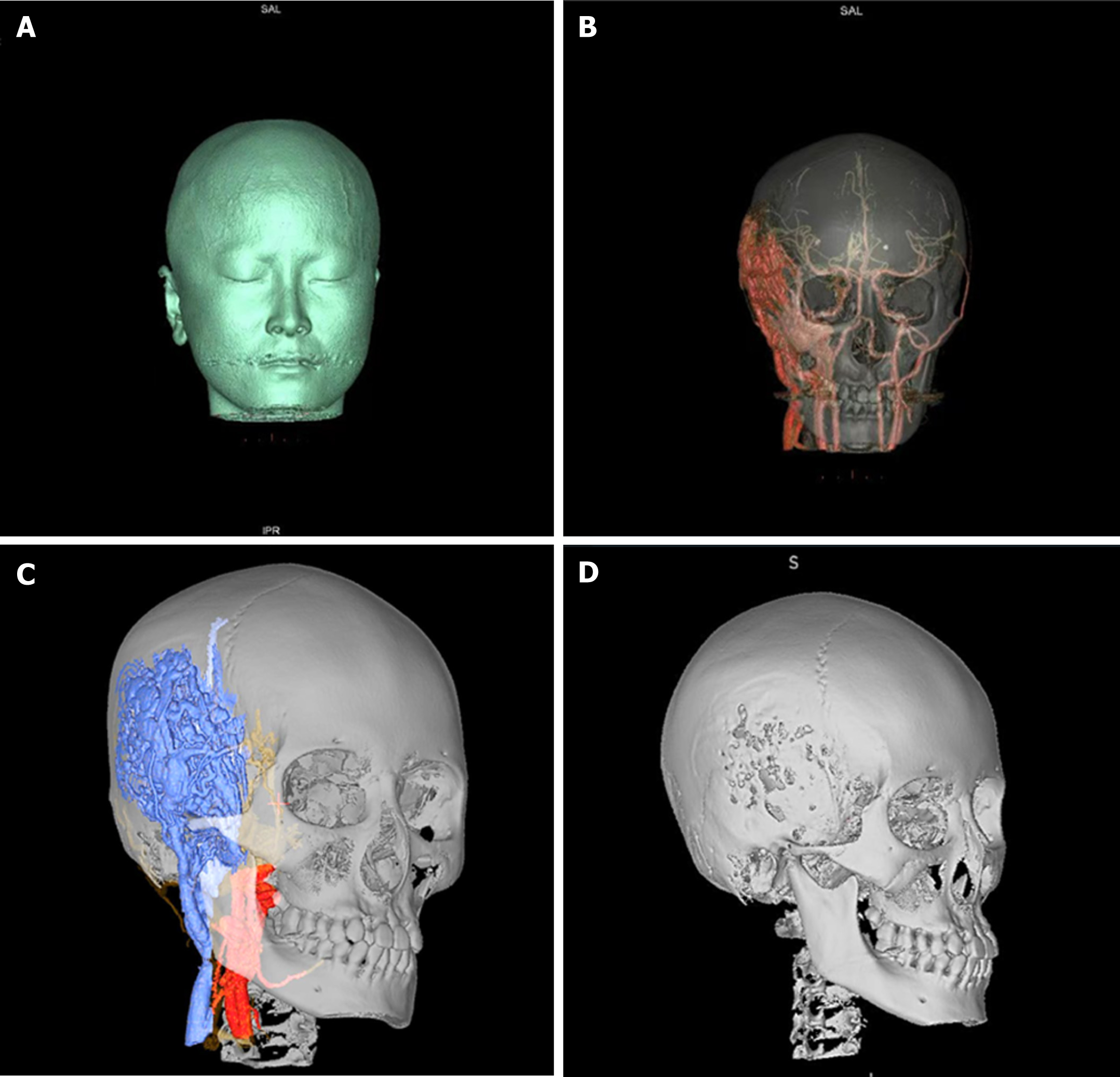
Figure 2 Trabecular vascular malformation under neuronavigation three dimensional reconstruction.
A: Mild protrusion of the patient's right temporal region that did not break down; B and C: Massive trabecular vascular malformation with extensive erosion of the right occlusal muscle and temporal bone; D: Worm-like changes of the skull.
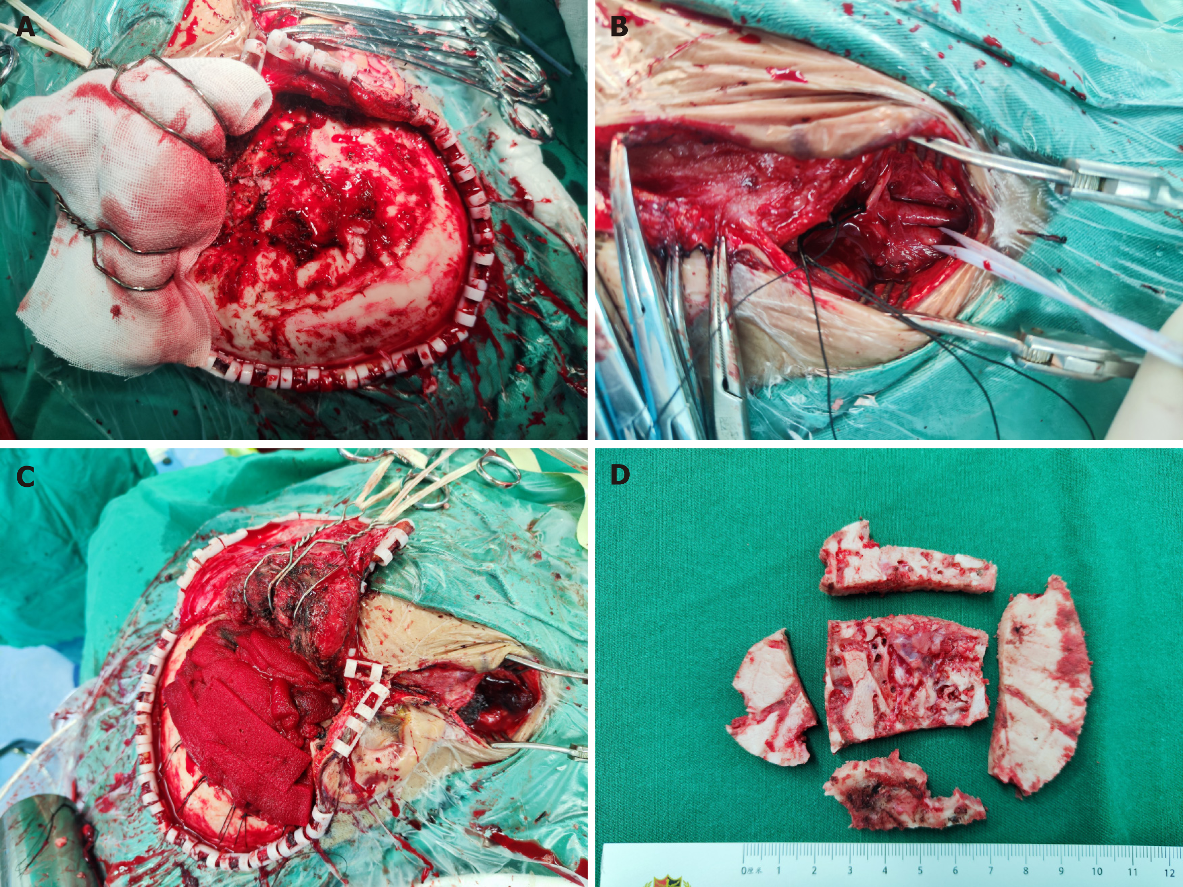
Figure 3 Surgical field images.
A: Raging cranial hemorrhage with massive bone wax to stop bleeding; B: Exposure of the right internal and external carotid artery and intraoperative ligation of the right external carotid artery first; C: Surgical incision on the right anterior cervical and right frontotemporal regions, and intraoperative excision of the eroded cranium; D: Intraoperative worm-like changes in the cranium.
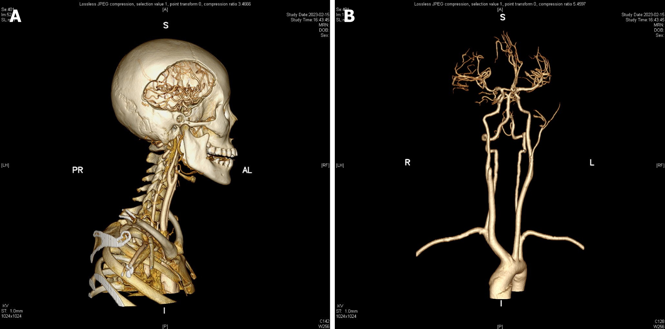
Figure 4 Postoperative review of the head and carotid artery computed tomography.
A: The eroded skull bone was removed to an approximate extent; B: The right external carotid artery was permanently ligated.
- Citation: Xie MC, Wang FX, Xu J. Giant vascular malformations invading the skull: A case report. World J Clin Cases 2024; 12(16): 2869-2875
- URL: https://www.wjgnet.com/2307-8960/full/v12/i16/2869.htm
- DOI: https://dx.doi.org/10.12998/wjcc.v12.i16.2869


