Copyright
©The Author(s) 2024.
World J Clin Cases. Jun 6, 2024; 12(16): 2862-2868
Published online Jun 6, 2024. doi: 10.12998/wjcc.v12.i16.2862
Published online Jun 6, 2024. doi: 10.12998/wjcc.v12.i16.2862
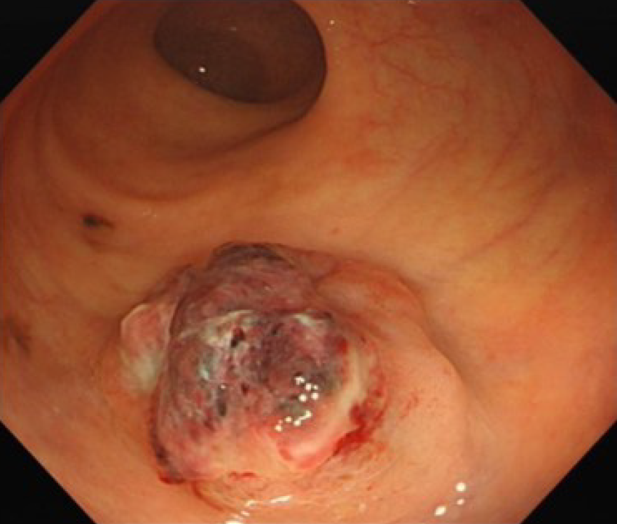
Figure 1 A bulging tumor of approximately 2.
2 cm × 2.0 cm with a purplish-red apex and clear boundaries was identified approximately 8 cm from the anus by colonoscopy.
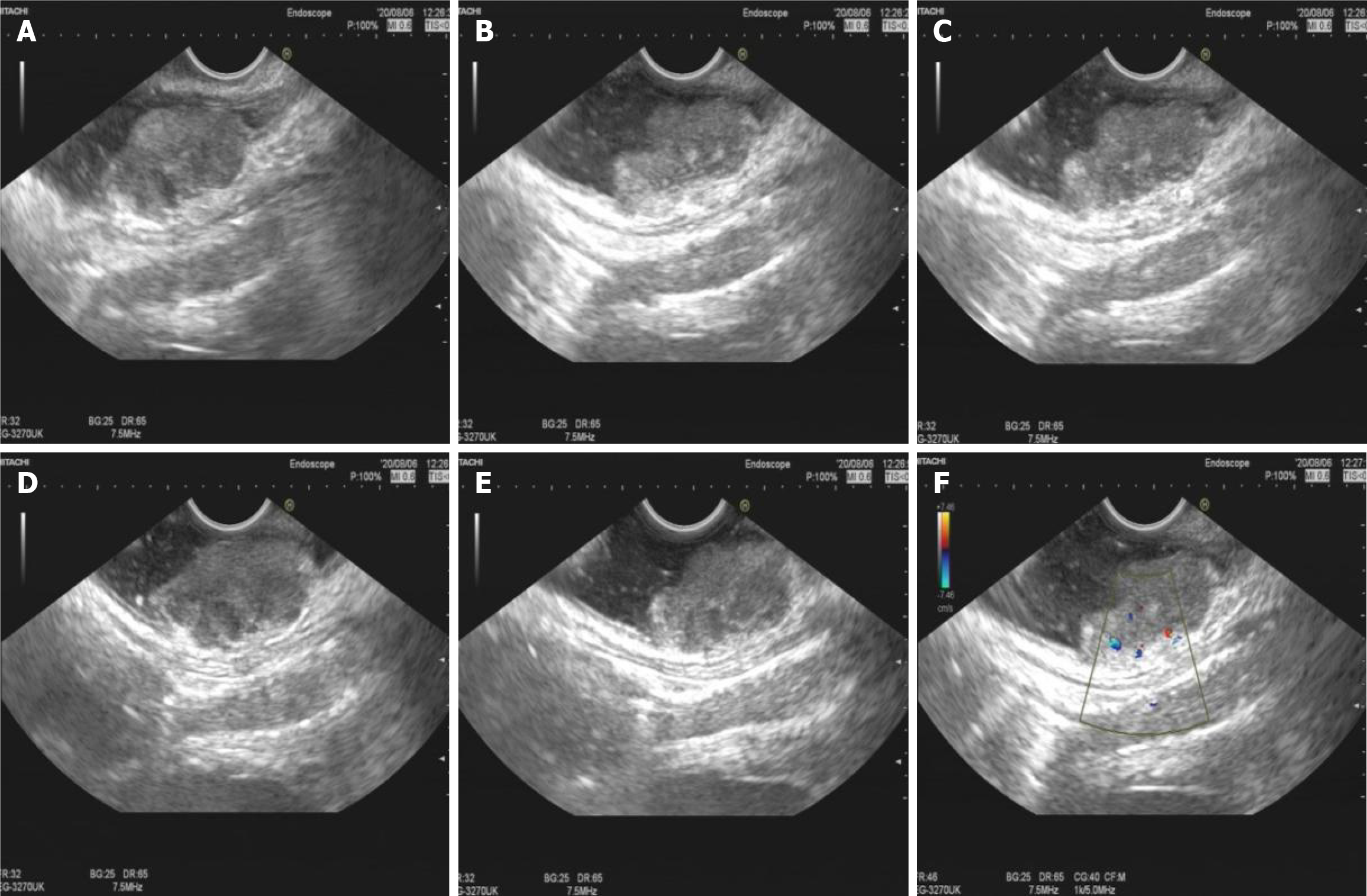
Figure 2 Endoscopic ultrasound image and color Doppler image.
A-E: Endoscopic ultrasound image showing a hypoechoic mucosal mass with involvement of the submucosal layer and heterogeneity of the internal echoes; F: Color Doppler showed localized blood with the intrinsic musculature still intact.
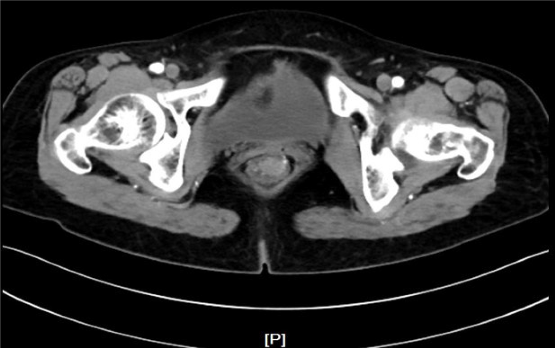
Figure 3 Nodular foci of enhancement in the rectum; abdominal contrast enhancement-computed tomography.
Figure 4 Hematoxylin-eosin staining and immunohistochemical staining.
A: Islands of malignant cells with round or oval nuclei of variable size and deep staining of the nuclei, accompanied by mucosal infiltration; hematoxylin and eosin staining (200-fold magnification); B: HMB-45 melanocyte marker, positive in tumor cells and negative in the remaining epithelium; Immunohistochemistry (IHC) (200-fold magnification); C: S100 melanocyte marker, positive for tumor cells, negative for remaining epithelial cells; IHC (200-fold magnification).
- Citation: Xiong ZE, Wei XX, Wang L, Xia C, Li ZY, Long C, Peng B, Wang T. Endoscopic ultrasound features of rectal melanoma: A case report and review of literature. World J Clin Cases 2024; 12(16): 2862-2868
- URL: https://www.wjgnet.com/2307-8960/full/v12/i16/2862.htm
- DOI: https://dx.doi.org/10.12998/wjcc.v12.i16.2862


